Elastofibroma.

Thompson LD.
Ear Nose Throat J. 2017 Apr-May;96(4-5):160.
FIRST PARAGRAPH: Elastofibroma is an ill-defined fibroelastic tumor-like condition that affects the neck and upper shoulders. There is a genetic predisposition, with multifocality suggesting an enzymatic defect that results from abnormal elastogenesis. Patients usually present in the 60- to 70-year range, with females affected much more often than males (5:1). Although perhaps due to reporting bias, there is an increased incidence in Japanese patients.
PubMed ID: 28489232
Article Size: <1 MB

New tumor entities in the 4th edition of the World Health Organization classification of head and neck tumors: Nasal cavity, paranasal sinuses and skull base

Thompson LDR, Franchi A.
Virchows Arch. 2018 Mar;472(3):315-330.
TheWorld Health Organization recently published the 4th edition of the Classification of Head and Neck Tumors, including several new entities, emerging entities, and significant updates to the classification and characterization of tumor and tumor-like lesions, specifically as it relates to nasal cavity, paranasal sinuses, and skull base in this overview. Of note, three new entities (NUT carcinoma, seromucinous hamartoma, biphenotypic sinonasal sarcoma,) were added to this section, while emerging entities (SMARCB1-deficient carcinoma and HPV-related carcinoma with adenoid cystic-like features) and several tumor-like entities (respiratory epithelial adenomatoid hamartoma, chondromesenchymal hamartoma) were included as provisional diagnoses or discussed in the setting of the differential diagnosis. The sinonasal tract houses a significant diversity of entities, but interestingly, the total number of entities has been significantly reduced by excluding tumor types if they did not occur exclusively or predominantly at this site or if they are discussed in detail elsewhere in the book. Refinements to nomenclature and criteria were provided to sinonasal papilloma, borderline soft tissue tumors, and neuroendocrine neoplasms. Overall, the new WHO classification reflects the state of current understanding for many relatively rare neoplasms, with this article highlighting the most significant changes.
PubMed ID: 28444451
Article Size: 6 MB

Neuroendocrine tumors of the larynx.

Thompson LD.
Ear Nose Throat J. 2016 Jul;95(7):262-6.
FIRST PARAGRAPH: Neuroendocrine neoplasms of the larynx encompass five separate tumors: paraganglioma, typical carcinoid, atypical carcinoid, small cell carcinoma, and large cell neuroendocrine carcinoma. Many synonyms are employed, with well-, moderately, and poorly differentiated neuroendocrine carcinoma commonly applied to the middle three tumors (World Health Organization preferred terminology). Paraganglioma is an exceedingly uncommon neuroendocrine tumor in the larynx, as is large cell neuroendocrine carcinoma, and therefore these will not be further discussed.
PubMed ID: 27434472
Article Size: <1 MB

Hailey-Hailey disease.

Thompson LD.
Ear Nose Throat J. 2016 Sep;95(9):370.
FIRST PARAGRAPH: Hailey-Hailey disease is a genodermatosis characterized by incomplete penetrance of an autosomal dominant disorder with defects in the ATP2C1 gene. Patients present with well-demarcated, erythematous plaques that often show crusting. The plaques may be vegetative, commonly associated with a distinct malodor. There may be associated burning or itching. While intertriginous areas are most commonly affected, the neck is also frequently involved. There is a natural remission and exacerbation cycle, with heat, moisture, and trauma the most common exacerbating factors. The lesions may be managed by corticosteroids or antibiotics, along with retinoids for severe disease.
PubMed ID: 27657314
Article Size: <1 MB

Secretory carcinoma.

Thompson LD.
Ear Nose Throat J. 2016 Dec;95(12):474-476.
FIRST PARAGRAPH: Secretory carcinoma, first described as mammary analogue secretory carcinoma (MASC), is a recently described, distinctive malignant salivary gland tumor that is quite similar to secretory breast carcinoma, defined by the t(12;15)(p13;q25) translocation resulting in an ETV6-NTRK3 fusion product. The vast majority of these tumors used to be included in the acinic cell carcinoma category.
PubMed ID: 27929593
Article Size: <1 MB

Thyroglossal duct cyst.

Thompson LD.
Ear Nose Throat J. 2017 Feb;96(2):54-55.
FIRST PARAGRAPH: Embryologically, the thyroglossal duct develops as the thyroid anlage descends from the foramen cecum at the base of the tongue to its final resting point in the pretracheal inferior midline neck. The duct usually involutes, but persistence may give rise to a clinical cyst. Thyroglossal duct remnant cysts (TGDCs) are one of the most common neck lesions seen clinically, showing a bimodal age distribution in the first and fifth decades, and identified in approximately 2.2/100,000 population at risk each year. While there is an equal sex distribution, males tend to predominate in pediatric patients while females predominate among adults.
PubMed ID: 28231361
Article Size: <1 MB

Ossifying fibroma of the maxilla and sinonasal tract: Case series

Liu JJ, Thompson LD, Janisiewicz AM, Shibuya TY, Keschner DB, Garg R, Lee JT.
Allergy Rhinol (Providence). 2017 Mar 1;8(1):32-36. doi: 10.2500/ar.2017.8.0190
BACKGROUND: Head and neck ossifying fibroma (OF) is a rare, benign, locally aggressive, fibro-osseous tumor. The mandible is the most common site of involvement, followed by the maxilla, and, less frequently, the sinonasal cavities, orbit, skull base, and calvarium. In this study, we aimed to expand our understanding of this entity by presenting a case series of OF that involved the maxilla and sinonasal tract.
METHODS: A multicenter retrospective review was performed on all the patients with a diagnosis of OF from 2004 to 2013. Data were collected with respect to age, sex, clinical presentation, treatment, and outcome.
RESULTS: A total of 13 patients were identified. The mean age was 37 years, with a female predominance (69%). The maxillary sinus was most frequently involved site (46%). Eighty-five percent underwent open surgical resection. After a mean follow-up time of 47.3 months, three patients (23%) developed recurrent disease; all of whom were treated with an open approach.
CONCLUSION: Of the maxilla and sinonasal tract is an uncommon clinicopathologic entity. Although a timely diagnosis may obviate the need for external approaches, open surgical resection is often still necessary for management of extensive lesions. Close follow-up and additional surgery may also be required to treat recurrent disease.
PubMed ID: 28381325
Article Size: <1 MB
 

Thyroglossal Duct Cyst Carcinomas in Pediatric Patients: Report of Two Cases with a Comprehensive Literature Review

Thompson LD, Herrera HB, Lau SK.
Head Neck Pathol. 2017 Dec;11(4):442-449.
Thyroglossal duct cyst (TGDC) carcinomas are rarely encountered in the pediatric population. The clinical behavior of these tumors in the pediatric setting is unclear and management is not well defined. Two cases of pediatric thyroglossal duct cyst carcinoma were identified in a review of all thyroglossal duct cysts diagnosed over a ten year period. These two cases were analyzed along with 57 cases of thyroglossal duct cyst carcinoma affecting patients less than 21 years of age compiled from the English literature. Fifty-nine patients (36 females, 23 males) aged 6–20 years (mean 15.0 years) were identified. All presented with an anterior midline neck mass, which was typically mobile and non-tender. The average tumor size was 2.6 cm. Histologically, all tumors were papillary thyroid carcinomas arising in a background of a thyroglossal duct cyst. The tumors exhibited a papillary, follicular, or mixed architecture with classical papillary thyroid carcinoma nuclear features. Soft tissue extension was present in 16 cases. All patients were managed surgically with a Sistrunk procedure, with additional thyroidectomy performed in 29 patients, combined with a lymph node dissection (n = 15), or a Sistrunk and lymph node dissection (n = 5). All patients were stage I at presentation, with 11 showing lymph node metastases. Postoperative radioactive iodine was employed in 20 patients. A papillary carcinoma of the thyroid gland was reported in four of the patients who had concurrent/subsequent thyroidectomies. Recurrences were reported in four patients, with distant metastases in one patient, who died of disease (13 months). Follow up data was available for 45 patients, with an overall mean follow-up of 54.5 months. All patients were alive, with the exception of one who died with disease. TGDC carcinomas in pediatric patients is associated with a good overall prognosis, best managed by Sistrunk procedure alone, with selected lymph node dissection if clinically indicated.
PubMed ID: 28293858
Article Size: 1.5 MB


Update From the 4th Edition of the World Health Organization Classification of Head and Neck Tumours: Tumours of the Ear.

Thompson LD.
Head Neck Pathol. 2017 Mar;11(1):78-87. doi: 10.1007/s12105-017-0790-5. Epub 2017 Feb 28.
The 2017 fourth edition of the World Health Organization Classification of Tumours, specifically as it relates to the ear (Chap. 9), has several changes. Importantly, the number of entities has been significantly reduced by omitting tumors or lesions if they do not occur exclusively or predominantly at this site or if they are discussed in detail elsewhere in the book. These entities include: embryonal rhabdomyosarcoma, osteoma, exostosis, angiolymphoid hyperplasia with eosinophilia, Schneiderian papilloma, inverted papilloma, lipoma of the internal auditory canal, hemangioma, hematolymphoid tumors, and secondary tumors. Paraganglioma was included in the neck chapter. New entries include otosclerosis and cholesteatoma, while refinements to nomenclature, classification and criteria were incorporated into the ceruminous gland tumors and epithelial tumors of the middle and inner ear. Specifically, the middle and inner ear were combined, as practical limitations of origin and imaging make a definitive separation artificial. The classification reflects the state of current understanding for these uncommon entities, with this update only highlighting selected entities that were the most significantly changed.
PubMed ID: 28247225
Article Size: 4.6 MB


Laryngeal Dysplasia, Squamous Cell Carcinoma, and Variants

Thompson, LDR.
Surg Pathol Clin. 2017 Mar;10(1):15-33.
Squamous cell carcinoma (SCC) is a malignant epithelial tumor showing evidence of squamous differentiation. It is the most common malignancy of the larynx, with several variants (verrucous, exophytic or papillary, spindle-cell, basaloid, acantholytic, adenosquamous) recognized, with well-established precursor lesions. Dysplasia is now separated into only low-grade and high-grade categories. Each SCC variant has unique cytomorphologic features and histologic differential diagnoses that are important to consider, as management and outcomes are different.
PubMed ID: 28153131
Article Size: 13 MB


Small round blue cell tumors of the sinonasal tract: a differential diagnosis approach.

Thompson LD.
Mod Pathol. 2017 Jan;30(s1):S1-S26. doi: 10.1038/modpathol.2016.119.
One of the most challenging diagnostic categories within tumors of the sinonasal tract is the small round blue cell tumors. Biopsies are usually small and limited, resulting in considerable diagnostic difficulty for practicing surgical pathologists. These tumors share several overlapping histologic and immunophenotypic findings while also showing considerable variation within and between cases. Specific tumor site of origin, imaging findings, and clinical findings must be combined with the histology and pertinent ancillary studies if the correct diagnosis is to be reached. Discrimination between neoplasms is critical as there are significant differences in therapy and overall outcome. It is important to have a well developed differential diagnosis for this category of tumors, where each of the diagnoses is considered, evaluated, and either confirmed or excluded from further consideration. In an undifferentiated tumor, showing a small round blue cell morphology, using the mnemonic ‘MR SLEEP’ helps to highlight tumors to consider: melanoma, mesenchymal chondrosarcoma, rhabdomyosarcoma, sinonasal undifferentiated carcinoma, squamous cell carcinoma (including NUT carcinoma), small cell osteosarcoma, lymphoma, esthesioneuroblastoma (olfactory neuroblastoma), Ewing sarcoma/primitive neuroectodermal tumor, pituitary adenoma, and plasmacytoma. A panel of pertinent immunohistochemistry studies, histochemistries and/or molecular tests should aid in reaching a diagnosis, especially when taking the pattern and intensity of reactions into consideration.
PubMed ID: 28060373
Article Size: 4 MB
 

Template for Reporting Results of Biomarker Testing of Specimens From Patients With Thyroid Carcinoma.

Chiosea S, Asa SL, Berman MA, Carty SE, Currence L, Hodak S, Nikiforov YE, Richardson MS, Seethala RR, Sholl LM, Thompson LD, Wenig BM, Worden F; Members of the Cancer Biomarker Reporting Committee, College of American Pathologists.
Arch Pathol Lab Med. 2017 Apr;141(4):559-563.
The College of American Pathologists offers these templates to assist pathologists in providing clinically useful and relevant information when reporting results of biomarker testing. The College regards the reporting elements in the templates as important elements of the biomarker test report, but the manner in which these elements are reported is at the discretion of each specific pathologist, taking into account clinician preferences, institutional policies, and individual practice.
The College developed these templates as educational tools to assist pathologists in the useful reporting of relevant information. It did not issue them for use in litigation, reimbursement, or other contexts. Nevertheless, the College recognizes that the templates might be used by hospitals, attorneys, payers, and others. The College cautions that use of the templates other than for their intended educational purpose may involve additional considerations that are beyond the scope of this document.
Completion of the template is the responsibility of the laboratory performing the biomarker testing and/or providing the interpretation. When both testing and interpretation are performed elsewhere (e.g., a reference laboratory), synoptic reporting of the results by the laboratory submitting the tissue for testing is also encouraged to ensure that all information is included in the patient’s medical record and thus readily available to the treating clinical team.
PubMed ID: 27681332
Article Size: 83 KB
 

Thyroglossal Duct Cyst Carcinomas: A Clinicopathologic Series of 22 Cases with Staging Recommendations

Thompson LD, Herrera HB, Lau SK.
Head Neck Pathol. 2017 Jun;11(2):175-185.
Carcinomas arising from thyroglossal duct remnant cysts (TGDCs) are rare, without well-defined management and staging criteria. All TGDCs (n = 685) diagnosed between 2005 and 2015 were retrospectively reviewed, with 22 carcinomas identified (3.2 % incidence). Twenty-two patients (17 females, 5 males), aged 12-64 years (mean 39.9 years; median 39 years) were identified. An anterior, superior midline neck mass was the presenting symptom in all patients. A cancer diagnosis [all papillary thyroid carcinoma (PTC)] was made after the Sistrunk procedure (SP), with a Bethesda Category V or VI classification preoperatively by fine needle aspiration in 5 of 12 cases tested. A SP was performed in all patients, with total thyroidectomy concurrently (n = 4) or subsequently (n = 12). A selected neck dissection was performed in 5 patients, with metastases found in 3. Of the patients who had a thyroidectomy, synchronous PTC was identified in 6 (thus, 6 of 22 patients had synchronous thyroid gland primaries). This supports an origin from extra-thyroidal remnants (cyst origin) rather than metastatic tumor from a thyroid gland primary. Follow-up radioactive iodine therapy was performed in 13 patients. Metastatic disease to local lymph nodes 57 months after presentation was seen in 1 patient, with all others alive and disease free (mean 3.8 years; range 0.4-10.8 years). The TGDCs ranged from 0.8 to 5 cm (mean 2.3 cm), while the PTCs ranged from 0.1 to 3.8 cm (mean 1.4 cm). All of the tumors were classical PTC, showing a sclerotic and infiltrative pattern, with a capsule present in 11. Lymphovascular invasion was detected in 11; margins were positive in 6. Using currently defined criteria, the patients were separated into AJCC stage group I (n = 21) or II (n = 1). However, if extension into the adipose tissue (n = 11), skeletal muscle (n = 10), or perineural/perivascular tissues (n = 10) were used to stage the patients, interpreted to represent the equivalent of “extrathyroidal extension” (n = 13) as defined for thyroid gland primaries, there would be 15 group I and 7 group III cases. All seven group III patients were ≥45 years. Three of four patients with lymph node metastasis also showed soft tissue extension. In conclusion, TGDC carcinomas (TGDCCa) are uncommon, with all classical PTC. For “microcarcinomas” (≤1 cm), conservative management can be used for patients <45 years (i.e., Sistrunk procedure only); for >1 cm tumors, and due to the high incidence of concurrent papillary carcinoma and higher stage at presentation in older patients, completion thyroidectomy is recommended for patients ≥45 years. Thus, even though a good prognosis can be expected for PTC developing in TGDCs, staging is advocated to more appropriately match therapeutic interventions.
PubMed ID: 27704385
Article Size: 2.5 MB


Myeloid sarcoma of the breast soft tissue following allogeneic bone marrow transplantation

Chou JL, Thompson LD.
J Case Rep Images Oncology 2016;2:71–73.
FIRST PARAGRAPH: A 47-year-old female presented with high-risk myelodysplastic syndrome (MDS) without detectable cytogenetic abnormality from the bone marrow specimen. Initially, she received azacytidine while awaiting bone marrow transplantation (BMT). After successful allogeneic BMT from a mismatched related donor, she received post-transplant care including management of transplant-related toxicity and skin manifestations of graft versus host disease (GVHD) in a designated BMT center. Subsequent bone marrow showed no evidence of MDS, and she is currently free from GVHD.
Article Size: <1 MB

Inter-Observer Variation in the Pathologic Identification of Extranodal Extension in Nodal Metastasis from Papillary Thyroid Carcinoma

Du E, Wenig BM, Su HK, Rowe ME, Haser GC, Asa SL, Baloch Z, Faquin WC, Fellegara G, Giordano T, Ghossein R, LiVolsi VA, Lloyd R, Mete O, Ozbek U, Rosai J, Suster S, Thompson LD, Turk AT, Urken ML.
Thyroid. 2016 Jun;26(6):816-9.
Background: Extranodal extension (ENE) in lymph node metastases has been shown to worsen the prognosis of papillary thyroid cancer (PTC). Despite the clinical significance of ENE, there are no stringent criteria for its microscopic diagnosis, and its identification is subject to inter-observer variability. The objective of this study was to determine the level of agreement among expert pathologists in the identification of ENE in PTC cases.
Methods: Eleven expert pathologists from the United States, Italy, and Canada were asked to review 61 scanned slides of representative permanent sections of PTC specimens from Mount Sinai Beth Israel Medical Center in New York. Each slide was evaluated for the presence of ENE. The pathologists were also asked to report the criteria they use to identify ENE.
Results: The overall strength of agreement in identifying ENE was only fair (j = 0.35), and the proportion of observed agreement was 0.68. The proportions of observed agreement for the identification of perinodal structures (fat, nerve, skeletal, and thick-walled vessel involvement) ranged from 0.61 to 0.997.
Conclusions: Overall agreement for the identification of ENE is poor. The lack of agreement results from both variation in pathologists’ identification of features and disagreement on the histologic criteria for ENE. This lack of concordance may help explain some of the discordant information regarding prognosis in clinical studies when this feature is identified.
PubMed ID: 27089928
Article Size: <1 MB
 

Subsets of Salivary Duct Carcinoma Defined by Morphologic Evidence of Pleomorphic Adenoma, PLAG1 or HMGA2 Rearrangements, and Common Genetic Alterations

Chiosea SI, Thompson LD, Weinreb I, Bauman JE, Mahaffey AM, Miller C, Ferris RL, Gooding WE.
Cancer. 2016 Oct 15;122(20):3136-3144. doi: 10.1002/cncr.30179. Epub 2016 Jul 5.
BACKGROUND: The authors hypothesized that histogenetic classification of salivary duct carcinoma (SDC) could account for de novo tumors and those with morphologic or molecular evidence (pleomorphic adenoma gene 1 [PLAG1], high-mobility group AT hook 2 [HMGA2] rearrangement, amplification) of pleomorphic adenoma (PA).
METHODS: SDCs (n = 66) were reviewed for morphologic evidence of PA. PLAG1 and HMGA2 alterations were detected by fluorescence in situ hybridization (FISH). PLAG1-positive tumors were tested by FISH for fibroblast growth factor receptor 1 (FGFR1) rearrangement. Thirty-nine tumors were analyzed using a commercial panel for mutations and copy number variations in 50 cancer-related genes.
RESULTS: On the basis of combined morphologic and molecular evidence of PA, 4 subsets of SDC emerged: 1) carcinomas with morphologic evidence of PA but intact PLAG1 and HMGA2 (n = 22); 2) carcinomas with PLAG1 alteration (n = 18) or 3) HMGA2 alteration (n = 12); and 4) de novo carcinomas, without morphologic or molecular evidence of PA (n = 14). The median disease-free survival was 37 months (95% confidence interval, 28.4-45.6 months). Disease-free survival and other clinicopathologic parameters did not differ for the subsets defined above. Combined Harvey rat sarcoma viral oncogene homolog/phosphatidylinositol-4,5-biphosphate 3-kinase, catalytic subunit α (HRAS/PIK3CA) mutations were observed predominantly in de novo carcinomas (5 of 8 vs 2 of 31 tumors; P = .035). Erb-B2 receptor tyrosine kinase 2 (ERBB2) copy number gain was not observed in de novo carcinomas (0 of 8 vs 12 of 31 tumors; P = .08). Tumor protein 53 (TP53) mutations were more common in SDC ex pleomorphic adenomas than in de novo carcinomas (17 of 31 vs 1 of 8 tumors; P = .033).
CONCLUSIONS: The genetic profile of SDC varies with the absence or presence of pre-existing PA and its cytogenetic signature. Most de novo SDCs harbor combined HRAS/PIK3CA mutations and no ERBB2 amplification.
PubMed ID: 27379604
Article Size: <1 MB


Cervical Lymph Node Metastasis in Adenoid Cystic Carcinoma of the Larynx: A Collective International Review

Coca-Pelaz A, Barnes L, Rinaldo A, Cardesa A, Shah JP, Rodrigo JP,, Suárez C,, Eloy JA, Bishop JA, Devaney KO, Thompson LD, Wenig BM, Strojan P, Hamoir M, Bradley PJ,, Gnepp DR,, Silver CE, Slootweg PJ, Triantafyllou A, Vander Poorten V,, Williams MD, Skálová A, Hellquist H, Teymoortash A, Medina JE, Robbins KT, Pitman KT, Kowalski LP, de Bree R, Mendenhall WM, Takes RP, Ferlito A.
Adv Ther. 2016 Apr;33(4):553-79.
Adenoid cystic carcinoma (AdCC) of the head and neck is a well-recognized pathologic entity that rarely occurs in the larynx. Although the 5-year locoregional control rates are high, distant metastasis has a tendency to appear more than 5 years post treatment. Because AdCC of the larynx is uncommon, it is difficult to standardize a treatment protocol. One of the controversial points is the decision whether or not to perform an elective neck dissection on these patients. Because there is contradictory information about this issue, we have critically reviewed the literature from 1912 to 2015 on all reported cases of AdCC of the larynx in order to clarify this issue. During the most recent period of our review (1991-2015) with a more exact diagnosis of the tumor histology, 142 cases were observed of AdCC of the larynx, of which 91 patients had data pertaining to lymph node status. Eleven of the 91 patients (12.1%) had nodal metastasis and, based on this low proportion of patients, routine elective neck dissection is therefore not recommended.
PubMed ID: 27084720
Article Size: <1 MB
 

Update on follicular variant of papillary thyroid carcinoma with an emphasis on new terminology: noninvasive follicular thyroid neoplasm with papillary-like nuclear features

Thompson, LDR.
Diagnostic Histopathology 2016.
The most common papillary thyroid carcinoma (PTC) variant is the follicular variant, representing w30% of all PTCs. The tumour is most common in middle aged (4th e 5th decades) women, who usually present with a single dominant nodule (about 3 cm). By definition, follicular architecture must be the dominant finding, while demonstrating the nuclear features of PTC. Papillary structures are <1% of volume, while necrosis, increased mitoses (>3/10 high power fields) and psammoma bodies are absent. The tumour category is divided into “encapsulated/well demarcated” and “invasive” types. The nuclear features include enlarged, elongated and overlapping nuclei; membrane irregularities (irregular contours, grooves and pseudoinclusions); chromatin clearing, margination and glassy nuclei. When the tumour is encapsulated/well demarcated without invasion, demonstrating the other inclusion and exclusion criteria, the new name of “Noninvasive Follicular Thyroid Neoplasm with Papillary-like Nuclear Features” (NIFTP) is used, a tumour that requires no additional treatment.
PubMed ID: n/a
Article Size: 8.4 MB

A Clinicopathologic Series of 685 Thyroglossal Duct Remnant Cysts

Thompson LD, Herrera HB, Lau SK.
Head Neck Pathol. 2016 Dec;10(4):465-474. Epub 2016 May 9.
The clinical features of thyroglossal duct remnant cysts (TGDC) have been well described, however the histopathologic aspects of these lesions have not been addressed in a detailed manner. In particular, there has been no large community practice based series evaluating TGDC histologically compared with management outcomes. A retrospective review of all TGDC diagnosed between 2005 and 2015 was performed. Six hundred eighty-five patients were identified (344 males; 341 females). Age at presentation was bimodal (first and fifth decades) and ranged from 0.8 to 87 years (mean 31.3 years). Males predominate in children (150:111); females in adults (230:194). Patients presented most frequently with a mobile midline neck mass in an infrahyoid location. An associated skin fistula (n = 67) was twice as common in pediatric as adult patients. The average cyst size was 2.4 cm (range 0.4-9.9 cm) by imaging studies and 2.6 cm (range 0.2-8.5 cm) by pathologic examination; pediatric patients had smaller cysts (mean 2.1 cm) than adults (mean 2.8 cm). Histologically, 257 (38 %) TGDC were lined by respiratory epithelium alone, 68 (10 %) squamous epithelium alone, 347 (51 %) exhibited both respiratory and squamous epithelium, and 13 (1 %) had no identifiable epithelial lining. Four hundred eighty-four (71 %) TGDC had associated thyroid gland tissue present within the cyst wall (n = 282), skeletal muscle (n = 71), adipose tissue (n = 34), or a combination of these sites (n = 97). The hyoid bone was identified in 647 (grossly and/or histologically), and absent in 38. Surgical management consisted of Sistrunk procedure (n = 647), cystectomy (n = 31), or thyroidectomy/thyroid lobectomy (n = 7). Treatment related complications were observed in 6 patients, which included vocal cord damage, seroma, and hematoma. Recurrences developed in 20 (3 %) patients, 14 of whom were managed initially by cystectomy. Papillary thyroid carcinoma was identified in 22 (3.2 %) TGDC. In summary, TGDC show a bimodal peak in the 1st and 5th decades, commonly presenting as a midline cervical lesion below the hyoid bone, associated with a skin fistula in 10 %. Histologically TGDC are most commonly lined by a combination of respiratory and squamous epithelium. Thyroid gland tissue is identified in 71 % of cases (0.45 cm mean size), although not limited to the cyst wall, but present in the surrounding soft tissues. Rare TGDC may harbor malignancy (3.2 %). TGDC are most effectively managed by Sistrunk procedure rather than excision, which carries low rates of complications (1 %) and recurrence (3 %).
PubMed ID: 27161104
Article Size: 2.7 MB


Ear and temporal bone meningioma

Thompson, LD.
Ear Nose Throat J. 2016 Apr-May;95(4-5):146.
FIRST PARAGRAPH: Meningiomas account for 30% of all intracranial neoplasms, but primary extracranial (ectopic) meningiomas of the ear and temporal bone are less common, accounting for about 10% of all ear and temporal bone tumors. Meningiomas are derived from the arachnoid cap cells, also called pacchionian bodies.
PubMed ID: 27140011
Article Size: <1 MB

Ninety-four cases of encapsulated follicular variant of papillary thyroid carcinoma: A name change to Noninvasive Follicular Thyroid Neoplasm with Papillary-like Nuclear Features would help prevent overtreatment

Thompson, LD.
Mod Pathol. 2016 Jul;29(7):698-707.
Encapsulated follicular variant of papillary thyroid carcinoma is a common thyroid gland cancer, with a highly indolent behavior. Recently, reclassification as a non-malignant neoplasm has been proposed. There is no comprehensive, community hospital based longitudinal evaluation of encapsulated follicular variant of papillary thyroid carcinoma. Ninety-four cases of encapsulated follicular variant of papillary thyroid carcinoma were identified in a review of all thyroid gland surgeries performed in 2002 within the Southern California Permanente Medical Group. All histology slides were reviewed and follow-up obtained. Seventy-five women and nineteen men, aged 20-80 years (mean 45.6 years), had a single (n=61), multiple (same lobe; n=20), or bilateral (n=13) tumor(s), ranging in size from 0.7 to 9.5 cm in diameter (mean 3.3 cm). Histologically, all cases demonstrated a well-formed tumor capsule, with capsular and/or lymphovascular invasion in 17 and no invasion in 77 cases. Lymph node metastases were not identified. The tumors had a follicular architecture, without necrosis or 43 mitoses/10 high-power fields (HPFs). Classical papillary thyroid carcinoma nuclear features were seen in at least three HPFs per 3mm of tumor diameter, including enlarged, elongated, crowded, and overlapping nuclei, irregular nuclear contours, nuclear grooves, and nuclear chromatin clearing. Lobectomy alone (n=41), thyroidectomy alone (n=34), or completion thyroidectomy (n=19) was the initial treatment combined with post-op radioablative iodine in 25 patients. All patients were without evidence of disease after a median follow-up of 11.8 years. Encapsulated follicular variant of papillary thyroid carcinoma showed benign behavior, supporting conservative surgery alone and reclassification of these tumors to Noninvasive Follicular Thyroid Neoplasm with Papillary-like Nuclear Features (NIFTP).
PubMed ID: 27102347
Article Size: <1 MB
 

Nomenclature Revision for Encapsulated Follicular Variant of Papillary Thyroid Carcinoma: A Paradigm Shift to Reduce Overtreatment of Indolent Tumors.

Nikiforov YE, Seethala RR, Tallini G, Baloch ZW, Basolo F, Thompson LD, Barletta JA, Wenig BM, Al Ghuzlan A, Kakudo K, Giordano TJ, Alves VA, Khanafshar E, Asa SL, El-Naggar AK, Gooding WE, Hodak SP, Lloyd RV, Maytal G, Mete O, Nikiforova MN, Nosé V, Papotti M, Poller DN, Sadow PM, Tischler AS, Tuttle RM, Wall KB, LiVolsi VA, Randolph GW, Ghossein RA.
JAMA Oncol. 2016 Aug 1;2(8):1023-9.
IMPORTANCE: Although growing evidence points to highly indolent behavior of encapsulated follicular variant of papillary thyroid carcinoma (EFVPTC), most patients with EFVPTC are treated as having conventional thyroid cancer.
OBJECTIVE: To evaluate clinical outcomes, refine diagnostic criteria, and develop a nomenclature that appropriately reflects the biological and clinical characteristics of EFVPTC.
DESIGN, SETTING, AND PARTICIPANTS: International, multidisciplinary, retrospective study of patients with thyroid nodules diagnosed as EFVPTC, including 109 patients with noninvasive EFVPTC observed for 10 to 26 years and 101 patients with invasive EFVPTC observed for 1 to 18 years. Review of digitized histologic slides collected at 13 sites in 5 countries by 24 thyroid pathologists from 7 countries. A series of teleconferences and a face-to-face conference were used to establish consensus diagnostic criteria and develop new nomenclature.
MAIN OUTCOMES AND MEASURES: Frequency of adverse outcomes, including death from disease, distant or locoregional metastases, and structural or biochemical recurrence, in patients with noninvasive and invasive EFVPTC diagnosed on the basis of a set of reproducible histopathologic criteria.
RESULTS: Consensus diagnostic criteria for EFVPTC were developed by 24 thyroid pathologists. All of the 109 patients with noninvasive EFVPTC (67 treated with only lobectomy, none received radioactive iodine ablation) were alive with no evidence of disease at final follow-up (median [range], 13 [10-26] years). An adverse event was seen in 12 of 101 (12%) of the cases of invasive EFVPTC, including 5 patients developing distant metastases, 2 of whom died of disease. Based on the outcome information for noninvasive EFVPTC, the name “noninvasive follicular thyroid neoplasm with papillary-like nuclear features” (NIFTP) was adopted. A simplified diagnostic nuclear scoring scheme was developed and validated, yielding a sensitivity of 98.6% (95% CI, 96.3%-99.4%), specificity of 90.1% (95% CI, 86.0%-93.1%), and overall classification accuracy of 94.3% (95% CI, 92.1%-96.0%) for NIFTP.
CONCLUSIONS AND RELEVANCE: Thyroid tumors currently diagnosed as noninvasive EFVPTC have a very low risk of adverse outcome and should be termed NIFTP. This reclassification will affect a large population of patients worldwide and result in a significant reduction in psychological and clinical consequences associated with the diagnosis of cancer.
PubMed ID: 27078145
 

Inter-Observer Variation in the Pathologic Identification of Minimal Extrathyroidal Extension in Papillary Thyroid Carcinoma.

Su HK, Wenig BM, Haser GC, Rowe ME, Asa SL, Baloch Z, Du E, Faquin WC, Fellegara G, Giordano T, Ghossein R, LiVolsi VA, Lloyd R, Mete O, Ozbek U, Rosai J, Suster S, Thompson LD, Turk AT, Urken ML.
Thyroid. 2016 Apr;26(4):512-7.
BACKGROUND: Extrathyroidal extension (ETE) is a significant prognostic factor in papillary thyroid carcinoma (PTC). Minimal extrathyroidal extension (mETE) is characterized by involvement of the sternothyroid muscle or perithyroid soft tissue, and is generally identified by light microscope examination. Patients with mETE, identified pathologically, are automatically upstaged to pT3. However, the prognostic implications of mETE have been a source of controversy in the literature. Moreover, there is also controversy surrounding the identification of mETE on pathological specimens. The objective of this study was to determine the level of agreement among expert pathologists in the identification of mETE in PTC cases.
METHODS: Eleven expert pathologists from the United States, Italy, and Canada were asked to perform a review of 69 scanned slides of representative permanent sections of PTC specimens. Each slide was evaluated for the presence of mETE. The pathologists were also asked to list the criteria they use to identify mETE.
RESULTS: The overall strength of agreement for identifying mETE was slight (κ = 0.14). Inter-pathologist agreement was best for perithyroidal skeletal muscle involvement (κ = 0.46, moderate agreement) and worst for invasion around thick-walled vascular structures (κ = 0.02, slight agreement). In addition, there was disagreement over the constellation of histologic features that are diagnostic for mETE, which affected overall agreement for diagnosing mETE.
CONCLUSIONS: Overall agreement for the identification of mETE is poor. Disagreement is a result of both variation in individual pathologists’ interpretations of specimens and disagreement on the histologic criteria for mETE. Thus, the utility of mETE in staging and treatment of PTC is brought into question. The lack of concordance may explain the apparent lack of agreement regarding the prognostic significance of this pathologic feature.
PubMed ID: 26953223
Article Size: <1 MB
 

Rhinosporidiosis.

Thompson LD.
Ear Nose Throat J. 2016 Mar;95(3):101..
FIRST PARAGRAPH: Rhinosporidium seeberi is the etiologic agent of a chronic, and usually painless, localized granulomatous infection of the mucous membranes of the sinonasal tract, conjunctiva, and urethra. Endemic in India and Sri Lanka, the disease is becoming more significant as a result of migration. R seeberi has not been definitively cultured but is thought to be a blue-green algae, with the infectious agent being a thick-walled sporangium containing endospores. It is passed to humans from animals (cats, free-grazing horses) or possibly fomites, identified in water or soil contaminated by waste.
PubMed ID: 26991216
Article Size: <1 MB

Cervical Lymph Node Metastasis in High-Grade Transformation of Head and Neck Adenoid Cystic Carcinoma: A Collective International Review.

Hellquist H, Skálová A, Barnes L, Cardesa A, Thompson LD, Triantafyllou A, Williams MD, Devaney KO, Gnepp DR,, Bishop JA, Wenig BM, Suárez C,, Rodrigo JP,, Coca-Pelaz A, Strojan P, Shah JP, Hamoir M, Bradley PJ,, Silver CE, Slootweg PJ, Vander Poorten V,, Teymoortash A, Medina JE, Robbins KT, Pitman KT, Kowalski LP, de Bree R, Mendenhall WM, Eloy JA, Takes RP, Rinaldo A, Ferlito A.
Adv Ther. 2016 Mar;33(3):357-68.
Adenoid cystic carcinoma (AdCC) is among the most common malignant tumors of the salivary glands. It is characterized by a prolonged clinical course, with frequent local recurrences, late onset of metastases and fatal outcome. High-grade transformation (HGT) is an uncommon phenomenon among salivary carcinomas and is associated with increased tumor aggressiveness. In AdCC with high-grade transformation (AdCC-HGT), the clinical course deviates from the natural history of AdCC. It tends to be accelerated, with a high propensity for lymph node metastasis. In order to shed light on this rare event and, in particular, on treatment implications, we undertook this review: searching for all published cases of AdCC-HGT. We conclude that it is mandatory to perform elective neck dissection in patients with AdCC-HGT, due to the high risk of lymph node metastases associated with transformation.
PubMed ID: 26895332
Article Size: 3 MB
 

Cervical lymph node metastasis in adenoid cystic carcinoma of oral cavity and oropharynx: A collective international review

Suárez C, Barnes L, Silver CE, Rodrigo JP , Shah JP, Triantafyllou A, Rinaldo A, Cardesa A, Pitman KT, Kowalski LP, Robbins KT , Hellquist H, Medina JE, de Bree R, Takes RP, Coca-Pelaz A , Bradley PJ, Gnepp DR, Teymoortash A, Strojan P, Mendenhall WM, Anderson Eloy J, Bishop JA, Devaney KO, Thompson LDR, Hamoir M, Slootweg PJ, Vander Poorten VV, Williams MD, Wenig BM, Skálová A, Ferlito A.
Auris Nasus Larynx. 2016 Oct;43(5):477-484.
The purpose of this study was to suggest general guidelines in the management of the N0 neck of oral cavity and oropharyngeal adenoid cystic carcinoma (AdCC) in order to improve the survival of these patients and/or reduce the risk of neck recurrences. The incidence of cervical node metastasis at diagnosis of head and neck AdCC is variable, and ranges between 3% and 16%. Metastasis to the cervical lymph nodes of intraoral and oropharyngeal AdCC varies from 2% to 43%, with the lower rates pertaining to palatal AdCC and the higher rates to base of the tongue. Neck node recurrence may happen after treatment in 0–14% of AdCC, is highly dependent on the extent of the treatment and is very rare in patients who have been treated with therapeutic or elective neck dissections, or elective neck irradiation. Lymph node involvement with or without extracapsular extension in AdCC has been shown in most reports to be independently associated with decreased overall and cause-specific survival, probably because lymph node involvement is a risk factor for subsequent distant metastasis. The overall rate of occult neck metastasis in patients with head and neck AdCC ranges from 15% to 44%, but occult neck metastasis from oral cavity and/or oropharynx seems to occur more frequently than from other locations, such as the sinonasal tract and major salivary glands. Nevertheless, the benefit of elective neck dissection (END) in AdCC is not comparable to that of squamous cell carcinoma, because the main cause of failure is not related to neck or local recurrence, but rather, to distant failure. Therefore, END should be considered in patients with a cN0 neck with AdCC in some high risk oral and oropharyngeal locations when postoperative RT is not planned, or the rare AdCC-high grade transformation.
PubMed ID: 27017314
Article Size: <1 MB
 

Diffuse hyperplasia of thyroid gland (Graves’ disease)

Thompson LDR.
Ear Nose Throat J. 2007 Nov;86(11):666-7.
FIRST PARAGRAPH: Widely known by its eponym Graves’ disease, diffuse hyperplasia of the thyroid gland is an autoimmune condition in which excess thyroid hormone production is unchecked by the normal feedback loop between the thyroid and the pituitary gland. The immune-system abnormality is mediated by antibodies to the thyrotropin receptor found on follicular epithelial cells. When the most specific antibody, known as thyroid-stimulating immunoglobulin (TSI), is bound to the thyrotropin receptor, the TSI mimics the action of pituitary thyrotropin and stimulates the follicular epithelium to produce hormone. The clinical effect of this autoimmune process is (1) thyrotoxicosis accompanied by diffuse thyroid enlargement and an infiltrative ophthalmopathy and (2) a spectrum of the systemic effects of excessive thyroid hormone production.
PubMed ID: 18225623
Article Size: <1 MB

Chondrodermatitis nodularis helicis.

Thompson LDR.
Ear Nose Throat J. 2007 Dec;86(12):734-5.
FIRST PARAGRAPH: Chondrodermatitis nodularis helicis (CDNH) is a non-neoplastic inflammatory and degenerative process of the external ear. It is characterized by necrobiotic changes in the dermis that extend down to the perichondrium; associated alterations are seen in the cartilaginous plate. The dermal injury is thought to be attributable to a combination of factors: local trauma, actinic damage, and the relatively tenuous vascularity of the auricle. The necrobiotic dermal collagen—and in some cases the cartilaginous matrix—is extruded through a crater-like defect in the epidermis; thus, CDNH is considered to be one of the transepidermal elimination disorders.
PubMed ID: 18217375
Article Size: <1 MB

Oral Traumatic Ulcer.

Thompson LD.
Ear Nose Throat J. 2011 Nov; 90(11):518-34.
FIRST PARAGRAPH: A traumatic ulcer is a chronic traumatic ulceration of the oral mucosa that shows unique histopathologic features. Also known as traumatic granuloma, eosinophilic granuloma, and Riga-Fede disease, this lesion is usually caused by some sort of mechanical injury. The most common causes include accidental trauma from biting, malposed teeth, and even sharp foodstuffs. However, self inflicted wounds caused by nocturnal clenching or tongue and lip biting, electrical and thermal injuries, hot foods or beverages, and even factitial injuries yield a similar finding. Ulceration of the ventral tongue as a result of tongue thrusting in infants with natal or neonatal teeth is referred to as Riga-Fede disease.
PubMed ID: 22109918
Article Size: <1 MB

Hotspot activating PRKD1 somatic mutations in polymorphous low-grade adenocarcinomas of the salivary glands

Weinreb I, Piscuoglio S, Martelotto LG, Waggott D, Ng CK, Perez-Ordonez B, Harding NJ, Alfaro J, Chu KC, Viale A, Fusco N, da Cruz Paula A, Marchio C, Sakr RA, Lim R, Thompson LD, Chiosea SI, Seethala RR, Skalova A, Stelow EB, Fonseca I, Assaad A, How C, Wang J, de Borja R, Chan-Seng-Yue M, Howlett CJ, Nichols AC, Wen YH, Katabi N, Buchner N, Mullen L, Kislinger T, Wouters BG, Liu FF, Norton L, McPherson JD, Rubin BP, Clarke BA, Weigelt B, Boutros PC, Reis-Filho JS.
Nat Genet. 2014;Nov;(46(11):1166-9.
Polymorphous low-grade adenocarcinoma (PLGA) is the second most frequent type of malignant tumor of the minor salivary glands. We identified PRKD1 hotspot mutations encoding p.Glu710Asp in 72.9% of PLGAs but not in other salivary gland tumors. Functional studies demonstrated that this kinase-activating alteration likely constitutes a driver of PLGA.
PubMed ID: 25240283
Article Size: 1 MB


Early Oral Tongue Squamous Cell Carcinoma: Sampling of Margins From Tumor Bed and Worse Local Control

Maxwell JH, Thompson LD, Brandwein-Gensler MS, Weiss BG, Canis M, Purgina B, Prabhu AV, Lai C, Shuai Y, Carroll WR, Morlandt A, Duvvuri U, Kim S, Johnson JT, Ferris RL, Seethala R, Chiosea S.
JAMA Otolaryngol Head Neck Surg. 2015 Dec 1;141(12):1104-10.
IMPORTANCE: Positive margins are associated with poor prognosis among patients with oral tongue squamous cell carcinoma (SCC). However, wide variation exists in the margin sampling technique.
OBJECTIVE: To determine the effect of the margin sampling technique on local recurrence (LR) in patients with stage I or II oral tongue SCC.
DESIGN, SETTING, AND PARTICIPANTS: A retrospective study was conducted from January 1, 1986, to December 31, 2012, in 5 tertiary care centers following tumor resection and elective neck dissection in 280 patients with pathologic (p)T1-2 pN0 oral tongue SCC. Analysis was conducted from June 1, 2013, to January 20, 2015.
INTERVENTIONS: In group 1 (n = 119), tumor bed margins were not sampled. In group 2 (n = 61), margins were examined from the glossectomy specimen, found to be positive or suboptimal, and revised with additional tumor bed margins. In group 3 (n = 100), margins were primarily sampled from the tumor bed without preceding examination of the glossectomy specimen. The margin status (both as a binary [positive vs negative] and continuous [distance to the margin in millimeters] variable) and other clinicopathologic parameters were compared across the 3 groups and correlated with LR.
MAIN OUTCOMES AND MEASURES: Local recurrence.
RESULTS: Age, sex, pT stage, lymphovascular or perineural invasion, and adjuvant radiation treatment were similar across the 3 groups. The probability of LR-free survival at 3 years was 0.9 and 0.8 in groups 1 and 3, respectively (P = .03). The frequency of positive glossectomy margins was lowest in group 1 (9 of 117 [7.7%]) compared with groups 2 and 3 (28 of 61 [45.9%] and 23 of 95 [24.2%], respectively) (P < .001). Even after excluding cases with positive margins, the median distance to the closest margin was significantly narrower in group 3 (2 mm) compared with group 1 (3 mm) (P = .008). The status (positive vs negative) of margins obtained from the glossectomy specimen correlated with LR (P = .007), while the status of tumor bed margins did not. The status of the tumor bed margin was 24% sensitive (95% CI, 16%-34%) and 92% specific (95% CI, 85%-97%) for detecting a positive glossectomy margin.
CONCLUSIONS AND RELEVANCE: The margin sampling technique affects local control in patients with oral tongue SCC. Reliance on margin sampling from the tumor bed is associated with worse local control, most likely owing to narrower margin clearance and greater incidence of positive margins. A resection specimen-based margin assessment is recommended.
PubMed ID: 26225798
Article Size: <1 MB


Update on Select Benign Mesenchymal and Meningothelial Sinonasal Tract Lesions

Thompson LD, Fanburg-Smith JC.
Head Neck Pathol. 2016 Mar;10(1):95-108.
Several benign and malignant mesenchymal and meningothelial lesions may preferentially affect or extend into the sinonasal tract. Glomangiopericytoma (GPC, formerly sinonasal-type hemangiopericytoma) is a specific tumor with a predilection to the sinonasal tract. Sinonasal tract polyps with stromal atypia (antrochoanal polyp) demonstrate unique histologic findings in the sinonasal tract. Juvenile nasopharyngeal angiofibroma (JNA) arises from specialized tissue in this location. Meningioma may develop as direct extension from its intracranial counterpart or as an ectopic tumor. Selected benign mesenchymal tumors may arise in the sinonasal tract and pose a unique differential diagnostic consideration, such as solitary fibrous tumor and GPC or lobular capillary hemangioma and JNA. Although benign and malignant vascular, fibrous, fatty, skeletal muscle, and nerve sheath tumors may occur in this location, this paper focuses on a highly select group of rare benign sinonasal tract tumors with their clinicopathological and molecular findings, and differential diagnosis.
PubMed ID: 26830398
Article Size: 4 MB


2016 SCPMG Thyroid NIFTP

Instructions for Validation of criteria for the new entity: Non-invasive Follicular Thyroid Neoplasm with Papillary-like Nuclear Features (NIFTP)

  1. Review these instructions and reply that you will be able to participate in the study in the time frame recommended
  2. Study the PowerPoint presentation, highlighting the features and criteria for the diagnosis
  3. Before beginning your review, ask Lester any questions, clarifications or concepts you may not clearly understand
  4. Please try to review the 30 cases in one or two seatings
  5. It will take about 30 minutes to look at the images, make a decision, and complete the appropriate line on the form
  6. The score you place (entry of 0 or 1) will be automatically added to create the 4th column
  7. Please provide your demographic data, which is only for Lester to use to avoid duplication, but also to allow the statistician to compare based on experience and volume of work
  8. Be able to complete this assignment before February 5, 2016. Return your completed Excel spreadsheet (see second tab of xls file; download below)
  9. Return Excel spreadsheet by email to Lester.D.Thompson@kp.org
  10. Aggregated results and comparison to the 26 endocrine pathologists will be provided as soon as the statistical evaluation is complete
  11. Data will be combined with academic pathologists from England (18-20 people), Japanese practicing pathologists (about 25), and the consensus conference group
  12. The information will be used to “validate” the criteria for reproducibility both between practicing pathologists and experts, but also against pathologists trained in different countries
Download Size: 12.4 MB
Download Size: 4.7 MB
Download Size: 23 KB Excel file

Pleomorphic sarcoma of the neck.

Thompson LD.
Ear Nose Throat J. 2015 Sep;94(9):376-7.
Pleomorphic sarcoma is an uncommon neoplasm in the head and neck now that refinements in diagnostic techniques have more accurately classified tumors that used to be placed in this category.

FIRST PARAGRAPH: ‘Pleomorphic sarcoma’ is the World Health Organization’s preferred term for malignant fibrous histiocytoma. This high-grade pleomorphic malignant mesenchymal neoplasm is a diagnosis of exclusion after other sarcomas and pleomorphic neoplasms have been excluded by histochemistry, immunohistochemistry, electron microscopy, and/or molecular evaluation. Most of these tumors arise de novo, but postradiation tumors are not uncommon. To be considered as a postradiation tumor, the tumor must be located in the radiation field and it must develop at least 3 years after radiation in an area that was free of tumor before radiation.

PubMed ID: 26401667
Article Size: <1 MB

Angiolymphoid hyperplasia with eosinophilia.

Thompson LD.
Ear Nose Throat J. 2015 Oct-Nov;94(10-11):443-4.

Angiolymphoid hyperplasia with eosinophilia (ALHE) is a benign vascular tumor that features immature blood vessels lined by epithelioid endothelial cells with a prominent inflammatory infiltrate, frequently showing a conspicuous eosinophil component.

FIRST PARAGRAPH: Angiolymphoid hyperplasia with eosinophilia (ALHE), sometimes called epithelioid hemangioma, is a benign vascular tumor. It features immature blood vessels lined by epithelioid endothelial cells with a prominent inflammatory infiltrate, frequently showing a conspicuous eosinophil component. There is controversy about whether this lesion is a reactive or benign neoplastic condition.

PubMed ID: 26535818
Article Size: <1 MB

Salivary gland adenoid cystic carcinoma.

Thompson LD.
Ear Nose Throat J. 2015 Jul;94(7):262-4.

Tumors are poorly circumscribed with an infiltrative border, including extracapsular extension beyond the salivary gland.

FIRST PARAGRAPH: Adenoid cystic carcinoma (ACC) is a malignant epithelial salivary gland tumor with myoepithelial and ductal differentiation. Salivary gland tumors account for only about 5% of all head and neck carcinomas, with ACC the fourth most common salivary gland malignancy. ACC has a 3:2 female-to-male ratio. Patients tend to be adults, with a peak clinical presentation in the sixth decade. The tumors are found most frequently in the parotid gland but are also found in the palate, tongue, lip, and other sites in the upper aerodigestive tract and the rest of the body.

PubMed ID: 26214665
Article Size: <1 MB

Salivary acinic cell carcinoma: reappraisal and update

Vander Poorten V, Triantafyllou A, Thompson LD, Bishop J, Hauben E, Hunt J, Skalova A, Stenman G, Takes RP, Gnepp DR, Hellquist H, Wenig B, Bell D, Rinaldo A, Ferlito A.
Eur Arch Otorhinolaryngol. 2016 Nov;273(11):3511-3531.
Epidemiologic and clinicopathologic features, therapeutic strategies, and prognosis for acinic cell carcinoma of the major and minor salivary glands are critically reviewed. We explore histopathologic, histochemical, electron microscopic and immunohistochemical aspects and discuss histologic grading, histogenesis, animal models, and genetic events. In the context of possible diagnostic difficulties, the relationship to mammary analog secretory carcinoma is probed and a classification is suggested. Areas of controversy or uncertainty, which may benefit from further investigations, are also highlighted.
PubMed ID: 26685679
Article Size: 5.22 MB
 

Angioleiomyoma of the Sinonasal Tract: Analysis of 16 Cases and Review of the Literature

Agaimy A, Michal M, Thompson LD, Michal M
Head Neck Pathol. 2015 Dec;9(4):463-73.
Angioleiomyoma (ALM; synonyms: angiomyoma, vascular leiomyoma) is an uncommon benign tumor of skin and subcutaneous tissue. Most arise in the extremities (90 %). Head and neck ALMs are uncommon (~10 % of all ALMs) and those arising beneath the sinonasal tract mucosa are very rare (<1 %) with 38 cases reported so far. We herein analyzed 16 cases identified from our routine and consultation files. Patients included seven females and nine males aged 25-82 years (mean 58; median 62). Symptoms were intermittent nasal obstruction, sinusitis, recurrent epistaxis, and a slow-growing mass. Fifteen lesions originated within different regions of the nasal cavity and one lesion was detected incidentally in an ethmoid sinus sample. Size range was 6-25 mm (mean 11). Histologically, all lesions were well circumscribed but non-encapsulated and most (12/16) were of the compact solid type superficially mimicking conventional leiomyoma but contained numerous compressed muscular veins. The remainder were of venous (2) and cavernous (2) type. Variable amounts of mature fat were observed in four cases (25 %). Atypia, necrosis, and mitotic activity were absent. Immunohistochemistry showed consistent expression of smooth muscle actin (12/12), h-caldesmon (9/9), muscle-specific actin (4/4), variable expression of desmin (11/14) and CD56 (4/6), and absence of HMB45 expression (0/11). The covering mucosa was ulcerated in 6 cases and showed squamous metaplasia in one case. There were no recurrences after local excision. Submucosal sinonasal ALMs are rare benign tumors similar to their reported cutaneous counterparts with frequent adipocytic differentiation. They should be distinguished from renal-type angiomyolipoma. Simple excision is curative.
PubMed ID: 26047608
Article Size: 3 MB
 

Low-Grade Fibromyxoid Sarcoma of the Head and Neck: A Clinicopathologic Series and Review of the Literature

Cowan ML, Thompson LD, Leon ME, Bishop JA.
Head Neck Pathol. 2016 Jun;10(2):161-6.
Low-grade fibromyxoid sarcoma (LGFMS) is a deceptively bland malignancy with potential for late recurrence and metastasis, which usually occurs in the deep soft tissues of the extremities and trunk. Most LGFMSs harbor a characteristic gene fusion of FUS-CREB3L2, and recently MUC4 immunostaining has been found to be highly sensitive and specific for the diagnosis. We present a dedicated series of head and neck LGFMS, including the first reported laryngeal case, as well as a review of reported head and neck cases. The surgical pathology archives of our three institutions were searched for cases of LGFMS arising within the head and neck, and four cases were identified. The H&E slides were reviewed, and immunohistochemistry were performed for pancytokeratin, p63, p40, EMA, S100 protein, β-catenin, actin, CD34, and MUC4. The patients were 6, 43, 45, and 73 years old (mean 41.8 years) and included three males and one female. The tumors were located in the posterior cervical spine, facial skin, mandible, and larynx. The tumors were treated with surgical excision, and all four had histologic features typical for LGFMS including alternating myxoid and fibrous areas with prominent curvilinear vasculature. All tumors were MUC4 positive (100%), 2/4 (50%) were p63 positive, 1/4 (25%) showed focal EMA positivity; all 4 were negative for pancytokeratin, p40, S100 protein, β-catenin, actin, and CD34. LGFMS is a low grade sarcoma that rarely develops in the head and neck. Due to its rarity, a pathologist may not consider LGFMS in the differential diagnosis of spindle cell neoplasms within the head and neck. Immunohistochemical staining is helpful, but stains should be selected carefully to avoid misdiagnosis.
PubMed ID: 26276044
Article Size: 1.4 MB
 

Head and Neck Pathology: A Volume in Foundations in Diagnostic Pathology Series

3rd Edition of Head and Neck Pathology: A Volume in Foundations in Diagnostic Pathology Series3rd Edition (current)

Lester D.R. Thompson, MD • Justin A. Bishop, MD

  • Hardcover: 784 pages
  • Publisher: Elsevier; 3rd Edition (February 15, 2018)
  • Language: English
  • ISBN-10: 0323479162
  • ISBN-13: 978-0323479165
  • Product Dimensions: 10.8 x 8.5 x 1.2 inches
  • 1st Edition published in 2006; 2nd Edition published in 2012

Read the Publisher’s description plus several editorial reviews

Buy the 3rd Edition

 
Head and Neck Pathology: A Volume in Foundations in Diagnostic Pathology Series 2nd Edition

2nd Edition

1st Edition of Head and Neck Pathology: A Volume in Foundations in Diagnostic Pathology Series

1st Edition

Chinese Version of Head and Neck Pathology: A Volume in Foundations in Diagnostic Pathology Series

1st Edition in Chinese

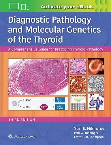
Diagnostic Pathology and Molecular Genetics of the Thyroid

Diagnostic Pathology and Molecular Genetics of the Thyroid (3rd Edition)3rd Edition (current)

Yuri E. Nikiforov, MD, PhD • Paul W. Biddinger, MD • Lester D.R. Thompson, MD FASCP FCAP

  • Hardcover: 480 pages
  • Publisher: Lippincott Williams & Wilkins; 3rd edition (December2018)
  • Language: English
  • ISBN-10: 1496396537
  • ISBN-13: 978-1496396532
  • Product Dimensions: 10.9 x 8.4

Buy this book

Read the Publisher’s description plus an editorial review

 
2nd Edition of Diagnostic Pathology and Molecular Genetics of the Thyroid
2nd Edition
1st Edition of Diagnostic Pathology and Molecular Genetics of the Thyroid
1st Edition

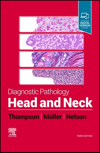
Diagnostic Pathology: Head and Neck

3rd Edition of Diagnostic Pathology: Head and Neck3rd Edition (current)

Lester D.R. Thompson, MD • Susan Müller, DMD • Brenda L. Nelson, DDS

Information:

  • Hardcover: 1217 pages
  • Publisher: Elsevier; 3rd edition (released 29 APR 2022)
  • Language: English
  • ISBN-10: 0323794068
  • ISBN-13: 978-0323794060
  • Product Dimensions: approx. 9.2 x 2.3 x 11.2 inches

Buy the 3rd Edition (current as of 4/2022)
Buy the 1st Edition in Spanish
 
Read the Publisher’s description plus various editorial reviews of this and previous editions


2nd Edition of Diagnostic Pathology: Head and Neck

2nd Edition

1st Edition of Diagnostic Pathology: Head and Neck

1st Edition

Diagnostic Pathology: Head and Neck in Spanish

1st Edition in Spanish

Protocol for the Examination of Specimens from Patients with Carcinomas of the of the Nasal Cavity and Paranasal Sinuses

College of American Pathologists (CAP)

Protocol applies to all invasive carcinomas of the nasal cavity and paranasal sinuses. Mucosal malignant melanoma is included. Lymphomas, neuroectodermal neoplasms, and sarcomas are not included.

Based on AJCC/UICC TNM, 7th edition
Protocol web posting date: October 2009
Protocol effective date: January 2010

Article Size: 1 MB

Protocol for the Examination of Specimens from Patients with Carcinomas of the Larynx

College of American Pathologists (CAP)
Protocol applies to all invasive carcinomas of the larynx, including supraglottis, glottis, and subglottis. Mucosal malignant melanoma is included. Lymphomas and sarcomas are not included.

Based on AJCC/UICC TNM, 7th edition
Protocol web posting date: October 2009
Protocol effective date: January 2010

Article Size: 1 MB

Squamous cell carcinoma variants of the head and neck.

Thompson LDR.
Curr Diag Pathol 2003 9, 384–396.
Variants of squamous cellcarcinoma (SCC) frequently arise within the mucosa of the upper aerodigestive tract, accounting for up to15% of SCCs in these areas. The most common variants include verrucous, exophytic or papillary, spindle-cell (sarcomatoid), basaloid and adenosquamous carcinoma. Each of these variants has a unique histomorphologic appearance, which raises a number of different differential diagnostic considerations, with the attendant clinically relevant management decision.
Verrucous squamous cell carcinoma has a broad border of pushing infiltration of a non-dysplastic squamous epithelium, essentially devoid of mitotic figures, displaying hyperkeratosis on elongated rete pegs. Papillary and exophytic SCC have a papillary or exophytic architecture, but have malignant cytologic features within the epithelium. Spindle-cell (sarcomatoid) carcinoma is an SCC blended with a spindle-cell morphology, frequently mimicking other mesenchymal tumours. Epithelial markers are often negative. Basaloid SCC is a high-grade SCC variant with small cells arranged in a palisaded architecture, with hyperchromatic nuclei and only focal areas of squamous differentiation. A denosquamous carcinoma is a rare variant, which is a composite of adenocarcinoma and squamous cell carcinoma, often with areas of transition.The cytomorphologic features are described in detail in an attempt to allow the general surgical pathologist to separate these variants of SCC in order to achieve appropriate clinical management.
PubMed ID: n/a
Article Size: 3 MB