Hotspot activating PRKD1 somatic mutations in polymorphous low-grade adenocarcinomas of the salivary glands

Weinreb I, Piscuoglio S, Martelotto LG, Waggott D, Ng CK, Perez-Ordonez B, Harding NJ, Alfaro J, Chu KC, Viale A, Fusco N, da Cruz Paula A, Marchio C, Sakr RA, Lim R, Thompson LD, Chiosea SI, Seethala RR, Skalova A, Stelow EB, Fonseca I, Assaad A, How C, Wang J, de Borja R, Chan-Seng-Yue M, Howlett CJ, Nichols AC, Wen YH, Katabi N, Buchner N, Mullen L, Kislinger T, Wouters BG, Liu FF, Norton L, McPherson JD, Rubin BP, Clarke BA, Weigelt B, Boutros PC, Reis-Filho JS.
Nat Genet. 2014;Nov;(46(11):1166-9.
Polymorphous low-grade adenocarcinoma (PLGA) is the second most frequent type of malignant tumor of the minor salivary glands. We identified PRKD1 hotspot mutations encoding p.Glu710Asp in 72.9% of PLGAs but not in other salivary gland tumors. Functional studies demonstrated that this kinase-activating alteration likely constitutes a driver of PLGA.
PubMed ID: 25240283
Article Size: 1 MB


Early Oral Tongue Squamous Cell Carcinoma: Sampling of Margins From Tumor Bed and Worse Local Control

Maxwell JH, Thompson LD, Brandwein-Gensler MS, Weiss BG, Canis M, Purgina B, Prabhu AV, Lai C, Shuai Y, Carroll WR, Morlandt A, Duvvuri U, Kim S, Johnson JT, Ferris RL, Seethala R, Chiosea S.
JAMA Otolaryngol Head Neck Surg. 2015 Dec 1;141(12):1104-10.
IMPORTANCE: Positive margins are associated with poor prognosis among patients with oral tongue squamous cell carcinoma (SCC). However, wide variation exists in the margin sampling technique.
OBJECTIVE: To determine the effect of the margin sampling technique on local recurrence (LR) in patients with stage I or II oral tongue SCC.
DESIGN, SETTING, AND PARTICIPANTS: A retrospective study was conducted from January 1, 1986, to December 31, 2012, in 5 tertiary care centers following tumor resection and elective neck dissection in 280 patients with pathologic (p)T1-2 pN0 oral tongue SCC. Analysis was conducted from June 1, 2013, to January 20, 2015.
INTERVENTIONS: In group 1 (n = 119), tumor bed margins were not sampled. In group 2 (n = 61), margins were examined from the glossectomy specimen, found to be positive or suboptimal, and revised with additional tumor bed margins. In group 3 (n = 100), margins were primarily sampled from the tumor bed without preceding examination of the glossectomy specimen. The margin status (both as a binary [positive vs negative] and continuous [distance to the margin in millimeters] variable) and other clinicopathologic parameters were compared across the 3 groups and correlated with LR.
MAIN OUTCOMES AND MEASURES: Local recurrence.
RESULTS: Age, sex, pT stage, lymphovascular or perineural invasion, and adjuvant radiation treatment were similar across the 3 groups. The probability of LR-free survival at 3 years was 0.9 and 0.8 in groups 1 and 3, respectively (P = .03). The frequency of positive glossectomy margins was lowest in group 1 (9 of 117 [7.7%]) compared with groups 2 and 3 (28 of 61 [45.9%] and 23 of 95 [24.2%], respectively) (P < .001). Even after excluding cases with positive margins, the median distance to the closest margin was significantly narrower in group 3 (2 mm) compared with group 1 (3 mm) (P = .008). The status (positive vs negative) of margins obtained from the glossectomy specimen correlated with LR (P = .007), while the status of tumor bed margins did not. The status of the tumor bed margin was 24% sensitive (95% CI, 16%-34%) and 92% specific (95% CI, 85%-97%) for detecting a positive glossectomy margin.
CONCLUSIONS AND RELEVANCE: The margin sampling technique affects local control in patients with oral tongue SCC. Reliance on margin sampling from the tumor bed is associated with worse local control, most likely owing to narrower margin clearance and greater incidence of positive margins. A resection specimen-based margin assessment is recommended.
PubMed ID: 26225798
Article Size: <1 MB


Update on Select Benign Mesenchymal and Meningothelial Sinonasal Tract Lesions

Thompson LD, Fanburg-Smith JC.
Head Neck Pathol. 2016 Mar;10(1):95-108.
Several benign and malignant mesenchymal and meningothelial lesions may preferentially affect or extend into the sinonasal tract. Glomangiopericytoma (GPC, formerly sinonasal-type hemangiopericytoma) is a specific tumor with a predilection to the sinonasal tract. Sinonasal tract polyps with stromal atypia (antrochoanal polyp) demonstrate unique histologic findings in the sinonasal tract. Juvenile nasopharyngeal angiofibroma (JNA) arises from specialized tissue in this location. Meningioma may develop as direct extension from its intracranial counterpart or as an ectopic tumor. Selected benign mesenchymal tumors may arise in the sinonasal tract and pose a unique differential diagnostic consideration, such as solitary fibrous tumor and GPC or lobular capillary hemangioma and JNA. Although benign and malignant vascular, fibrous, fatty, skeletal muscle, and nerve sheath tumors may occur in this location, this paper focuses on a highly select group of rare benign sinonasal tract tumors with their clinicopathological and molecular findings, and differential diagnosis.
PubMed ID: 26830398
Article Size: 4 MB


2016 SCPMG Thyroid NIFTP

Instructions for Validation of criteria for the new entity: Non-invasive Follicular Thyroid Neoplasm with Papillary-like Nuclear Features (NIFTP)

  1. Review these instructions and reply that you will be able to participate in the study in the time frame recommended
  2. Study the PowerPoint presentation, highlighting the features and criteria for the diagnosis
  3. Before beginning your review, ask Lester any questions, clarifications or concepts you may not clearly understand
  4. Please try to review the 30 cases in one or two seatings
  5. It will take about 30 minutes to look at the images, make a decision, and complete the appropriate line on the form
  6. The score you place (entry of 0 or 1) will be automatically added to create the 4th column
  7. Please provide your demographic data, which is only for Lester to use to avoid duplication, but also to allow the statistician to compare based on experience and volume of work
  8. Be able to complete this assignment before February 5, 2016. Return your completed Excel spreadsheet (see second tab of xls file; download below)
  9. Return Excel spreadsheet by email to Lester.D.Thompson@kp.org
  10. Aggregated results and comparison to the 26 endocrine pathologists will be provided as soon as the statistical evaluation is complete
  11. Data will be combined with academic pathologists from England (18-20 people), Japanese practicing pathologists (about 25), and the consensus conference group
  12. The information will be used to “validate” the criteria for reproducibility both between practicing pathologists and experts, but also against pathologists trained in different countries
Download Size: 12.4 MB
Download Size: 4.7 MB
Download Size: 23 KB Excel file

Pleomorphic sarcoma of the neck.

Thompson LD.
Ear Nose Throat J. 2015 Sep;94(9):376-7.
Pleomorphic sarcoma is an uncommon neoplasm in the head and neck now that refinements in diagnostic techniques have more accurately classified tumors that used to be placed in this category.

FIRST PARAGRAPH: ‘Pleomorphic sarcoma’ is the World Health Organization’s preferred term for malignant fibrous histiocytoma. This high-grade pleomorphic malignant mesenchymal neoplasm is a diagnosis of exclusion after other sarcomas and pleomorphic neoplasms have been excluded by histochemistry, immunohistochemistry, electron microscopy, and/or molecular evaluation. Most of these tumors arise de novo, but postradiation tumors are not uncommon. To be considered as a postradiation tumor, the tumor must be located in the radiation field and it must develop at least 3 years after radiation in an area that was free of tumor before radiation.

PubMed ID: 26401667
Article Size: <1 MB

Angiolymphoid hyperplasia with eosinophilia.

Thompson LD.
Ear Nose Throat J. 2015 Oct-Nov;94(10-11):443-4.

Angiolymphoid hyperplasia with eosinophilia (ALHE) is a benign vascular tumor that features immature blood vessels lined by epithelioid endothelial cells with a prominent inflammatory infiltrate, frequently showing a conspicuous eosinophil component.

FIRST PARAGRAPH: Angiolymphoid hyperplasia with eosinophilia (ALHE), sometimes called epithelioid hemangioma, is a benign vascular tumor. It features immature blood vessels lined by epithelioid endothelial cells with a prominent inflammatory infiltrate, frequently showing a conspicuous eosinophil component. There is controversy about whether this lesion is a reactive or benign neoplastic condition.

PubMed ID: 26535818
Article Size: <1 MB

Salivary gland adenoid cystic carcinoma.

Thompson LD.
Ear Nose Throat J. 2015 Jul;94(7):262-4.

Tumors are poorly circumscribed with an infiltrative border, including extracapsular extension beyond the salivary gland.

FIRST PARAGRAPH: Adenoid cystic carcinoma (ACC) is a malignant epithelial salivary gland tumor with myoepithelial and ductal differentiation. Salivary gland tumors account for only about 5% of all head and neck carcinomas, with ACC the fourth most common salivary gland malignancy. ACC has a 3:2 female-to-male ratio. Patients tend to be adults, with a peak clinical presentation in the sixth decade. The tumors are found most frequently in the parotid gland but are also found in the palate, tongue, lip, and other sites in the upper aerodigestive tract and the rest of the body.

PubMed ID: 26214665
Article Size: <1 MB

Salivary acinic cell carcinoma: reappraisal and update

Vander Poorten V, Triantafyllou A, Thompson LD, Bishop J, Hauben E, Hunt J, Skalova A, Stenman G, Takes RP, Gnepp DR, Hellquist H, Wenig B, Bell D, Rinaldo A, Ferlito A.
Eur Arch Otorhinolaryngol. 2016 Nov;273(11):3511-3531.
Epidemiologic and clinicopathologic features, therapeutic strategies, and prognosis for acinic cell carcinoma of the major and minor salivary glands are critically reviewed. We explore histopathologic, histochemical, electron microscopic and immunohistochemical aspects and discuss histologic grading, histogenesis, animal models, and genetic events. In the context of possible diagnostic difficulties, the relationship to mammary analog secretory carcinoma is probed and a classification is suggested. Areas of controversy or uncertainty, which may benefit from further investigations, are also highlighted.
PubMed ID: 26685679
Article Size: 5.22 MB
 

Angioleiomyoma of the Sinonasal Tract: Analysis of 16 Cases and Review of the Literature

Agaimy A, Michal M, Thompson LD, Michal M
Head Neck Pathol. 2015 Dec;9(4):463-73.
Angioleiomyoma (ALM; synonyms: angiomyoma, vascular leiomyoma) is an uncommon benign tumor of skin and subcutaneous tissue. Most arise in the extremities (90 %). Head and neck ALMs are uncommon (~10 % of all ALMs) and those arising beneath the sinonasal tract mucosa are very rare (<1 %) with 38 cases reported so far. We herein analyzed 16 cases identified from our routine and consultation files. Patients included seven females and nine males aged 25-82 years (mean 58; median 62). Symptoms were intermittent nasal obstruction, sinusitis, recurrent epistaxis, and a slow-growing mass. Fifteen lesions originated within different regions of the nasal cavity and one lesion was detected incidentally in an ethmoid sinus sample. Size range was 6-25 mm (mean 11). Histologically, all lesions were well circumscribed but non-encapsulated and most (12/16) were of the compact solid type superficially mimicking conventional leiomyoma but contained numerous compressed muscular veins. The remainder were of venous (2) and cavernous (2) type. Variable amounts of mature fat were observed in four cases (25 %). Atypia, necrosis, and mitotic activity were absent. Immunohistochemistry showed consistent expression of smooth muscle actin (12/12), h-caldesmon (9/9), muscle-specific actin (4/4), variable expression of desmin (11/14) and CD56 (4/6), and absence of HMB45 expression (0/11). The covering mucosa was ulcerated in 6 cases and showed squamous metaplasia in one case. There were no recurrences after local excision. Submucosal sinonasal ALMs are rare benign tumors similar to their reported cutaneous counterparts with frequent adipocytic differentiation. They should be distinguished from renal-type angiomyolipoma. Simple excision is curative.
PubMed ID: 26047608
Article Size: 3 MB
 

Low-Grade Fibromyxoid Sarcoma of the Head and Neck: A Clinicopathologic Series and Review of the Literature

Cowan ML, Thompson LD, Leon ME, Bishop JA.
Head Neck Pathol. 2016 Jun;10(2):161-6.
Low-grade fibromyxoid sarcoma (LGFMS) is a deceptively bland malignancy with potential for late recurrence and metastasis, which usually occurs in the deep soft tissues of the extremities and trunk. Most LGFMSs harbor a characteristic gene fusion of FUS-CREB3L2, and recently MUC4 immunostaining has been found to be highly sensitive and specific for the diagnosis. We present a dedicated series of head and neck LGFMS, including the first reported laryngeal case, as well as a review of reported head and neck cases. The surgical pathology archives of our three institutions were searched for cases of LGFMS arising within the head and neck, and four cases were identified. The H&E slides were reviewed, and immunohistochemistry were performed for pancytokeratin, p63, p40, EMA, S100 protein, β-catenin, actin, CD34, and MUC4. The patients were 6, 43, 45, and 73 years old (mean 41.8 years) and included three males and one female. The tumors were located in the posterior cervical spine, facial skin, mandible, and larynx. The tumors were treated with surgical excision, and all four had histologic features typical for LGFMS including alternating myxoid and fibrous areas with prominent curvilinear vasculature. All tumors were MUC4 positive (100%), 2/4 (50%) were p63 positive, 1/4 (25%) showed focal EMA positivity; all 4 were negative for pancytokeratin, p40, S100 protein, β-catenin, actin, and CD34. LGFMS is a low grade sarcoma that rarely develops in the head and neck. Due to its rarity, a pathologist may not consider LGFMS in the differential diagnosis of spindle cell neoplasms within the head and neck. Immunohistochemical staining is helpful, but stains should be selected carefully to avoid misdiagnosis.
PubMed ID: 26276044
Article Size: 1.4 MB
 

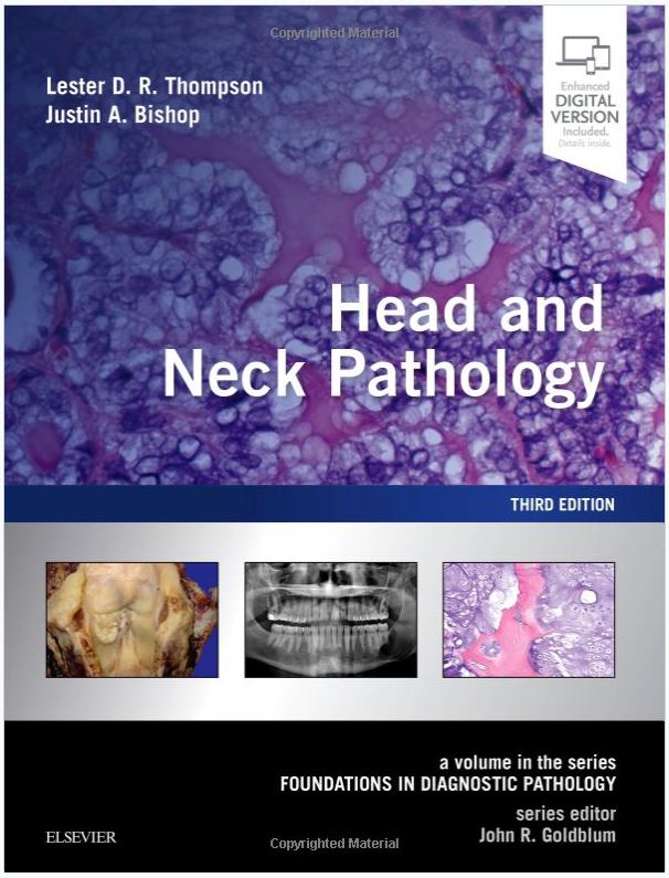
Head and Neck Pathology: A Volume in Foundations in Diagnostic Pathology Series

3rd Edition of Head and Neck Pathology: A Volume in Foundations in Diagnostic Pathology Series3rd Edition (current)

Lester D.R. Thompson, MD • Justin A. Bishop, MD

  • Hardcover: 784 pages
  • Publisher: Elsevier; 3rd Edition (February 15, 2018)
  • Language: English
  • ISBN-10: 0323479162
  • ISBN-13: 978-0323479165
  • 1st Edition published in 2006; 2nd Edition published in 2012

Read the Publisher’s description plus several editorial reviews

Buy the 3rd Edition

 
Head and Neck Pathology: A Volume in Foundations in Diagnostic Pathology Series 2nd Edition

2nd Edition

1st Edition of Head and Neck Pathology: A Volume in Foundations in Diagnostic Pathology Series

1st Edition

Chinese Version of Head and Neck Pathology: A Volume in Foundations in Diagnostic Pathology Series

1st Edition in Chinese

Diagnostic Pathology: Head and Neck

3rd Edition of Diagnostic Pathology: Head and Neck3rd Edition (current)

Lester D.R. Thompson, MD • Susan Müller, DMD • Brenda L. Nelson, DDS

  • Hardcover: 1217 pages
  • Publisher: Elsevier; 3rd edition (released 29 APR 2022)
  • Language: English
  • ISBN-10: 0323794068
  • ISBN-13: 978-0323794060

Buy the 3rd Edition (current as of 4/2022)

 
Read the Publisher’s description plus various editorial reviews of this and previous editions


2nd Edition of Diagnostic Pathology: Head and Neck

2nd Edition

1st Edition of Diagnostic Pathology: Head and Neck

1st Edition

Diagnostic Pathology: Head and Neck in Spanish

1st Edition in Spanish

Protocol for the Examination of Specimens from Patients with Carcinomas of the of the Nasal Cavity and Paranasal Sinuses

College of American Pathologists (CAP)

Protocol applies to all invasive carcinomas of the nasal cavity and paranasal sinuses. Mucosal malignant melanoma is included. Lymphomas, neuroectodermal neoplasms, and sarcomas are not included.

Based on AJCC/UICC TNM, 7th edition
Protocol web posting date: October 2009
Protocol effective date: January 2010

Article Size: 1 MB

Protocol for the Examination of Specimens from Patients with Carcinomas of the Larynx

College of American Pathologists (CAP)
Protocol applies to all invasive carcinomas of the larynx, including supraglottis, glottis, and subglottis. Mucosal malignant melanoma is included. Lymphomas and sarcomas are not included.

Based on AJCC/UICC TNM, 7th edition
Protocol web posting date: October 2009
Protocol effective date: January 2010

Article Size: 1 MB

Squamous cell carcinoma variants of the head and neck.

Thompson LDR.
Curr Diag Pathol 2003 9, 384–396.
Variants of squamous cellcarcinoma (SCC) frequently arise within the mucosa of the upper aerodigestive tract, accounting for up to15% of SCCs in these areas. The most common variants include verrucous, exophytic or papillary, spindle-cell (sarcomatoid), basaloid and adenosquamous carcinoma. Each of these variants has a unique histomorphologic appearance, which raises a number of different differential diagnostic considerations, with the attendant clinically relevant management decision.
Verrucous squamous cell carcinoma has a broad border of pushing infiltration of a non-dysplastic squamous epithelium, essentially devoid of mitotic figures, displaying hyperkeratosis on elongated rete pegs. Papillary and exophytic SCC have a papillary or exophytic architecture, but have malignant cytologic features within the epithelium. Spindle-cell (sarcomatoid) carcinoma is an SCC blended with a spindle-cell morphology, frequently mimicking other mesenchymal tumours. Epithelial markers are often negative. Basaloid SCC is a high-grade SCC variant with small cells arranged in a palisaded architecture, with hyperchromatic nuclei and only focal areas of squamous differentiation. A denosquamous carcinoma is a rare variant, which is a composite of adenocarcinoma and squamous cell carcinoma, often with areas of transition.The cytomorphologic features are described in detail in an attempt to allow the general surgical pathologist to separate these variants of SCC in order to achieve appropriate clinical management.
PubMed ID: n/a
Article Size: 3 MB

Sinonasal carcinomas.

Thompson LDR.
Curr Diag Pathol 2006 12, 40–53.
Malignant neoplasms of the sinonasal tract encompass a wide variety of epithelial, lymphoid and mesenchymal tumours. The separation and classification of epithelial or neuroepithelial tumours is sometimes challenging, especially when treatment and prognosis are different. Squamous cell carcinoma, keratinizing or non-keratinizing and, usually, the poorly differentiated type need to be separated from sinonasal undifferentiated carcinoma, lymphoepithelial carcinoma, neuroendocrine carcinoma and olfactory neuroblastoma. Whereas melanoma and lymphoma are also included in the broad differential, along with primitive neuroectodermal tumours and rhabdomyosarcomas, the focus of this commentary will be to present the major clinical, radiographical, histological, immunohistochemical, ultrastructural and molecular features which allow for separation of the principle mucosal epithelial neoplasms of the sinonasal tract.
PubMed ID: n/a
Article Size: 2 MB

Polymorphous low grade adenocarcinoma.

Thompson LDR.
Pathol Case Rev 2004;9:259-263.
Polymorphous low-grade adenocarcinomas are minor salivary gland neoplasms with a predilection for intraoral sites. Women are affected twice as frequently as men, and generally present in the fifth to sixth decade of life with a painless ihaoral mass. The palatal mass is, on average, about 2 cm in greatest dimension. The tumors are submucosal, identified below an intact mucosa as a well-circumscribed although unencapsulated mass. The tumor is characterized by a polymorphous growth pattern, with individual tumors demonstrating multiple patterns, including solid, ductal-tubular, cribriform, trabecular, and single-file growth. Neurotropism is common, frequently forming a central nidus around which a “targetoid” pattern is formed. The neoplastic cells are isomorphic, containing round to oval vesicular nuclei with small nucleoli. Mitotic activity and necrosis are inconspicuous. There is frequently a slate gray-blue stroma separating the tumor cells. Immunohistochemical analysis demonstrates reactivity with cytokeratin, vimentin, S-100 protein, CD117, glial fibrillary acidic protein, and actin. Bcl-2 is overexpressed and there is generally a low proliferation index as determined by Ki-67 reactions. The tumor must be separated from pleomorphic adenoma (benign mixed tumor) and adenoid cystic carcinoma. Complete surgical excision will yield a more than 95% 10-year survival, although persistence or recurrence can emerge often in about 10% of patients more than 10 years later.
PubMed ID: n/a
Article Size: 4.9 MB

Pheochromocytoma.

Thompson LDR.
Pathol Case Rev 2005;10 (5): 243-25.
Pheochromocytomas are tumors arising from the chromaffin cells of the adrenal medulla. They are equivalent to paragangliomas in other anatomic sites. They are uncommon neoplasms and most are sporadic, although 10% are described in syndromes, another 10% are bilateral (usually syndrome associated), and about 10% are malignant. They develop in both genders and peak in the fourth to fifth decades, although familial tumors occur at a younger age. Patients present clinically with the pharmacologic effects of excess catecholamines, manifested by episodic, postural, paroxysmal and/or labile hypertension, headaches, diaphoresis, palpitations, chest pain, and anxiety. Radiographic studies with nucleotide scans are often diagnostic. Malignant pheochromocytomas have historically required the presence of metastatic tumor to confirm the diagnosis of a malignant neoplasm. However, a series of histologic features, when used in conjunction with laboratory findings, radiographic findings, macroscopic features, and immunohistochemical results, can help to prospectively diagnose malignant heochromocytoma. These features include invasion, large nests or diffuse growth (loss of Zellballen architecture), focal or confluent necrosis, high cellularity, tumor cell spindling, cellular monotony, increased mitotic figures, atypical mitotic figures, profound nuclear pleomorphism, and hyperchromasia. Overall, the patient prognosis for benign pheochromocytoma is excellent, although that for malignant pheochromocytoma is intermediate, with surgery achieving the best clinical result of about 75% 5-year survival. Separation from benign pheochromocytoma and adrenal cortical neoplasms is important as the management is different.
PubMed ID: n/a
Article Size: 4 MB

Parotid Gland Nodular Fasciitis: A Clinicopathologic Series of 12 Cases with a Review of 18 Cases from the Literature.

Gibson TC, Bishop JA, Thompson LD.
Head Neck Pathol. 2015 Sep;9(3):334-44.
Nodular fasciitis (NF), very uncommon in the parotid gland, is a benign myofibroblastic proliferation that may be mistaken for other neoplastic proliferations. The mass-like clinical presentation and histologic features result in frequent misclassification, resulting in inappropriate clinical management. There are only a few reported cases in the English literature. Cases within the files of the authors’ institutions (retrospective) confined to the parotid gland were compared to cases reported in the English literature (Medline 1966–2014). The patients included five females and seven males, aged 11–70 years (mean 45.2 years). All patients presented with a mass lesion, present on average 1.9 months, without a documented history of trauma. The lesions were 0.7–5.2 cm (mean 2.2 cm). Seven patients had fine needle aspiration. The majority of the lesions were circumscribed (n = 9), composed of spindle-shaped to stellate myofibroblasts (MF) arranged in a storiform growth pattern, juxtaposed to hypocellular myxoid tissue-culture-like areas with extravasation of erythrocytes. Dense, keloid-like collagen (n = 7) and occasional giant cells were seen (n = 6). Mitotic figures (without atypical forms) were readily identifiable (mean 4/10 HPFs). By immunohistochemical staining, the MF were reactive with vimentin, actins, and calponin, while the histiocytes were reactive with CD68. All patients had surgical excision. One patient developed local recurrence (12 months later). All were alive and disease free at last follow-up, with a mean 133 months of follow-up. The principle differential diagnoses include fibrosarcoma, fibromatosis, pleomorphic adenoma, myoepithelioma, neurofibroma, schwannoma, solitary fibrous tumor, leiomyoma, fibrous histiocytoma and myxoma. NF of the parotid gland occurs in middle-aged patients who present with a mass (mean 2.2 cm) in the parotid gland of short duration (1.9 months). FNA misinterpretation frequently leads to excision. Separation from myoepithelial and mesenchymal lesions affecting the parotid gland results in appropriate management.
PubMed ID: 25472697
Article Size: 3.7 MB


Parathyroid carcinoma occurring in multiple endocrine neoplasia type 1.

Kodroff KS, Lieber C, Thompson LDR.
Endocr Pract. 1999 Nov-Dec;5(6):347-349.
OBJECTIVE: To describe a patient with multiple endocrine neoplasia type I (MEN-I) in whom parathyroid carcinoma developed.
METHODS: A case report is presented, including detailed history, laboratory findings, and pathologic features.
RESULTS: A 49-year-old man with a known history of MEN-I syndrome had hypercalcemia and profoundly increased parathyroid hormone levels. Seven years earlier, he had been diagnosed with a gastrinoma, prolactinoma, and primary hyperparathyroidism. A neck exploration at that time consisted of resection of a right inferior parathyroid gland and parathyroid tissue in the thyroid gland as well as biopsies of two left-sided glands, after which microscopic examination revealed parathyroid hyperplasia and his serum calcium levels normalized. On reexploration of the neck, resection revealed pathologic findings consistent with parathyroid carcinoma.
CONCLUSION: Parathyroid carcinoma has rarely been reported in the setting of MEN-I. This case illustrates the need for near-complete resection of parathyroid tissue in the patient with MEN-I. Subtotal or total parathyroidectomy in conjunction with forearm autotransplantation should be performed, not simply to prevent recurrence of benign disease but also to prevent the potential development of carcinoma over time.
PubMed ID: n/a
Article Size: 1.5 MB
 

Incisional or core biopsies of salivary gland tumours: how far should we go?

Nelson BL, Thompson LDR.
Diagnostic Pathology Volume 18:9 September 2012 pages 358-365.
Trends in the evaluation of salivary gland masses have changed as imaging studies have improved and sampling techniques have evolved over the past several decades. Whether clinically palpable or detected by imaging studies, salivary gland masses have been readily evaluated by fine needle aspiration. More recently, imaging guided core-needle biopsy has been employed with mixed results. The literature on these techniques is reviewed and analyzed with particular attention to tissue adequacy and diagnostic accuracy. Comparison is made using selected case presentations to highlight the advantages and disadvantages of establishing a diagnosis when core-needle biopsy is utilized. Core-needle biopsy of salivary gland tumours may be a useful first diagnostic approach as long as the limitations of the procedure are well understood and managed.
PubMed ID: n/a
Article Size: <1 MB

Exophytic and papillary squamous cell carcinoma of the larynx.

Kardon DE, Thompson LDR.
Pathol Case Rev 2000;5(4):196-199.
Conventional squamous cell carcinoma (SCC) of the larynx presents as a largely flat or ulcerated lesion and is usually categorized histologically as in situ, superficially invasive, or deeply invasive. Papillary and exophytic squamous cell carcinomas of the larynx represent uncommon but distinct subtypes of SCC. As implied by their designations, these tumor types show predominant papillary or exophytic growth in the form of finger-like or filiform and broad-based bulbous fronds, respectively. Both tumor types are composed of cytologically malignant squamous epithelium. The following case illustrates the clinical and pathologic features of papillary squamous cell carcinoma and emphasizes the importance of distinguishing this entity from conventional squamous cell carcinoma of the larynx.
PubMed ID: n/a
Article Size: 2.5 MB

Endoscopic Management of Recurrent Anterior Skull Base Schwannoma

Lee J, Thompson L, Garg R, Keschner D, Shibuya T.
International Journal of Otolaryngology and Head & Neck Surgery, Vol. 2 No. 2, 2013, pp. 52-56.
Objectives: Sinonasal schwannomas account for less than 4% of head and neck schwannomas, with the primary treatment modality being surgical excision via external approaches. The aim of this report is to present a rare case of recurrent schwannoma of the ethmoid cavity involving the anterior skull base which was successfully managed with endoscopic resection. Study Design: Case report and review of the literature. Methods: The clinical presentation, radiographic features, histopathologic characteristics, surgical approach, and patient outcome were examined in the context of a literature review. Results: A 43-year-old woman presented with a 9-month history of left facial pain and pressure. She had a prior history of sinonasal schwannoma excision with cerebrospinal fluid (CSF) leak repair via bifrontal craniotomy in 2007. Magnetic resonance imaging (MRI) and nasal endoscopy revealed a left ethmoid mass measuring 2.2 cm × 2.7 cm × 2.4 cm abutting the anterior skull base. The tumor was completely removed using a transnasal endoscopic approach, and the anterior skull base reconstructed with tensor fascia lata graft. Histology of the specimen showed schwannoma, and there has been no evidence of tumor recurrence nor CSF leak after 24 months of follow-up. Conclusion: With continual advances in surgical technique and instrumentation, sinonasal schwannomas have become increasingly more amenable to endoscopic resection even in the case of recurrence and skull base involvement.
PubMed ID: n/a
Article Size: >1 MB

BOOK REVIEW: ‘Biopsy Interpretation of the Upper Aerodigestive Tract and Ear’ by Edward B. Stelow, Stacey E. Mills.

Thompson LD.
Am J Surg Pathol. 2013 Oct;37(10):1632.
FIRST PARAGRAPH OF REVIEW: A fan of the Biopsy Interpretation series, I was pleased to review a new title, although without reviewing the first edition. As advertised, ‘recent advances in our understanding of aerodigestive tract pathology,’ are presented in a userfriendly manner, incorporating selected and targeted use of ancillary techniques to help reach a diagnosis, although the reader is reminded to focus on the standard hematoxylin and eosin-stained material first and foremost. In fact, the entire chapter on hematopathology only has a single immunohistochemistry image, which is normally a field run amok by mmunohistochemistry stains. Always forward thinking, and utilizing all resources available to reach a diagnosis, the book itself will be as helpful as any of the supporting ancillary studies.
PubMed ID: n/a
Article Size: <1 MB

Clinicopathologic and Immunophenotypic Characterization of 25 Cases of Acinic Cell Carcinoma with High-Grade Transformation

Thompson LD, Aslam MN, Stall JN, Udager AM, Chiosea S, McHugh JB
Head Neck Pathol. 2016 Jun;10(2):152-60.
Acinic cell carcinoma (AiCC) with high-grade transformation is a rare variant of AiCC composed of both a conventional low-grade (LG) AiCC and a separate highgrade (HG) component. We describe here, the clinicopathologic and immunohistochemical features of 25 cases diagnosed between 1990 and 2015. Available tissue was analyzed and compared with a cohort of pure LG AiCC for the morphologic and immunophenotypic profile. Incidence was higher in females (1.8:1) than males with an overall mean age at presentation of 63.2 years. All tumors occurred in the parotid gland including 76 % with facial nerve trunk and branches involvement. Most patients were treated with extensive resection and adjuvant therapy. Local recurrence or distant metastasis occurred in most patients, with 72.7 % dead with disease (mean 2.9 years) and 3 patients alive with disease (mean 2.4 years). The majority of the tumors were composed of a LG microcystic AiCC and a HG component consisting of invasive lobules of undifferentiated cells with predominantly solid, cribriform, and glandular patterns. Acinic differentiation was still present in HG areas but aggressive features such as perineural invasion (76 %), lymphovascular invasion (62 %), positive margins (72 %), high mitotic rate, atypical mitoses and/or comedonecrosis (86 %) were easily identified. Compared to the pure LG AiCC, the cases with HG transformation showed significantly increased expression of cyclin-D1, p53 and Ki-67. Most HG areas of AiCC expressed membranous b-catenin (92 %) and were negative for p63 (three cases were focally positive), S100, SMA, androgen, and estrogen receptors. DOG1 expression was present in all LG AiCC tested with retained expression in 91 % of cases with HG transformation, supporting acinic differentiation in the HG foci. Recognition of AiCC with high-grade transformation is imperative as more aggressive clinical management is warranted.
PubMed ID: 26245749
Article Size: 6 MB


Evaluation of PAX2 and PAX8 Expression in Salivary Gland Neoplasms.

Butler RT, Alderman MA, Thompson LD, McHugh JB.
Head Neck Pathol. 2015 Mar;9(1):47-50.
PAX2 and PAX8 are transcription factors involved in embryogenesis that have been utilized as immunohistochemical indicators of tumor origin. Specifically, PAX2 is a marker of neoplasms of renal and müllerian origin, while PAX8 is expressed by renal, müllerian, and thyroid tumors. While studies examining these transcription factors in a variety of tumors have been published, data regarding their expression in salivary gland neoplasms are limited. The goal of this study was to assess expression of PAX2 and PAX8 in a large cohort of salivary gland tumors. Utilizing tissue microarrays, samples of normal salivary glands (n = 68) and benign and malignant salivary gland neoplasms (n = 442) were evaluated for nuclear immunoreactivity with PAX2 and PAX8. No expression was observed with either marker in the normal salivary glands, and PAX8 was negative in all neoplasms. Focal expression of PAX2 was observed in one example each of oncocytoma and acinic cell carcinoma. These results indicate that evaluation of PAX2 and/or PAX8 expression would be valuable in differentiating primary salivary gland tumors from metastases known to express PAX2 and/or PAX8.
PubMed ID: 24771139
Article Size: <1 MB
 

Salivary Duct Carcinoma: The Predominance of Apocrine Morphology, Prevalence of Histologic Variants, and Androgen Receptor Expression

Williams L, Thompson LD, Seethala RR, Weinreb I, Assaad AM, Tuluc M, Ud Din N, Purgina B, Lai C, Griffith CC, Chiosea SI.
Am J Surg Pathol. 2015 May;39(5):705-13.
Salivary duct carcinoma (SDC) is a prototypic aggressive salivary gland carcinoma. Our aim is to determine the prevalence of histologic variants (micropapillary, basal-like) and androgen receptor (AR) expression in a large multi-institutional series of SDC. AR status was determined by immunohistochemistry (IHC). Most SDCs were characterized by an apocrine phenotype and AR expression. Cases with a nonapocrine phenotype and AR-negative status were studied by additional IHC and fluorescence in situ hybridization for ETV6 or MYB/NFIB. The diagnosis of SDC was confirmed in 187 of 199 (94%) cases. Variant morphologies were identified in 12 cases: micropapillary (n=6), sarcomatoid (n=3), mucinous (n=2), and basal-like (n=1). AR IHC was performed in 183 cases, of which 179 (97.8%) showed AR expression. On the basis of morphologic appearance and results of additional studies, 12 cases were reclassified as squamous cell carcinoma (SCC) (n=4), epithelial-myoepithelial carcinoma with high-grade transformation (HGT) (n=2), myoepithelial carcinoma (n=2), mammary analogue secretory carcinoma, high grade (ETV6 translocated; n=1), adenoid cystic carcinoma with HGT (n=1), acinic cell carcinoma with HGT (n=1), and adenosquamous carcinoma (n=1). AR-negative SDC is extremely rare, and the majority of such cases are more accurately classified as other entities. HGTs of other salivary carcinomas and squamous cell carcinoma are the most common mimics of SDC. SDCs with variant morphologies still show at least a minor component of conventional apocrine appearance. Thus, apocrine morphology defines SDC.
PubMed ID: 25871467
Article Size: 1 MB


Adenoid cystic carcinoma of the head and neck — An update

Coca-Pelaz A, Rodrigo JP, Bradley PJ, Vander Poorten V, Triantafyllou A, Hunt JL, Strojan P, Rinaldo A, Haigentz M Jr, Takes RP, Mondin V, Teymoortash A, Thompson LDR, Ferlito A.
Oral Oncol. 2015 Jul;51(7):652-661.
This article provides an update on the current understanding of adenoid cystic carcinoma of the head and neck, including a review of its epidemiology, clinical behavior, pathology, molecular biology, diagnostic workup, treatment and prognosis. Adenoid cystic carcinoma is an uncommon salivary gland tumor that may arise in a wide variety of anatomical sites in the head and neck, often with an advanced stage at diagnosis. The clinical course is characterized by very late recurrences; consequently, clinical follow-up should extend at least >15 years. The optimal treatment is generally considered to be surgery with postoperative radiotherapy to optimize local disease control. Much effort has been invested into understanding the tumor’s molecular biological processes, aiming to identify patients at high risk of recurrence, in hopes that they could benefit from other, still unproven treatment modalities such as chemotherapy or biological therapy.
PubMed ID: 25943783
Article Size: 2.6 MB
 

Rhabdomyoblastic Differentiation in Head and Neck Malignancies Other Than Rhabdomyosarcoma.

Bishop JA, Thompson LD, Cardesa A, Barnes L, Lewis JS Jr, Triantafyllou A, Hellquist H, Stenman G, Hunt JL, Williams MD, Slootweg PJ, Devaney KO, Gnepp DR, Wenig BM, Rinaldo A, Ferlito A.
Head Neck Pathol. 2015 Dec;9(4):507-18.
Rhabdomyosarcoma is a relatively common soft tissue sarcoma that frequently affects children and adolescents and may involve the head and neck. Rhabdomyosarcoma is defined by skeletal muscle differentiation which can be suggested by routine histology and confirmed by immunohistochemistry for the skeletal muscle-specific markers myogenin or myoD1. At the same time, it must be remembered that when it comes to head and neck malignancies, skeletal muscle differentiation is not limited to rhabdomyosarcoma. A lack of awareness of this phenomenon could lead to misdiagnosis and, subsequently, inappropriate therapeutic interventions. This review focuses on malignant neoplasms of the head and neck other than rhabdomyosarcoma that may exhibit rhabdomyoblastic differentiation, with an emphasis on strategies to resolve the diagnostic dilemmas these tumors may present. Axiomatically, no primary central nervous system tumors will be discussed.
PubMed ID: 25757816
Article Size: 3.8 MB


Cherubism.

Thompson LD.
Ear Nose Throat J. 2015 Jan;94(1):22-4.
FIRST PARAGRAPH: Cherubism is an autosomal-dominant inherited disease with variable expression. It is characterized by a progressive, painless, and symmetric expansion of the jaws. The disease is caused by a point mutation in the SH3BP2 gene (chromosome 4p16.3), which leads to dysregulation of the Msx-1 gene; this gene is involved in regulating mesenchymal interaction in craniofacial morphogenesis.
PubMed ID: 25606831
Article Size: <1 MB

Sox10 — A Marker for Not Only Schwannian and Melanocytic Neoplasms But Also Myoepithelial Cell Tumors of Soft Tissue: A Systematic Analysis of 5134 Tumors

Miettinen M, McCue PA, Sarlomo-Rikala M, Biernat W, Czapiewski P, Kopczynski J, Thompson LD, Lasota J, Wang Z, Fetsch JF.
Am J Surg Pathol. 2015 Jun;39(6):826-35.
Sox10 transcription factor is expressed in schwannian and melanocytic lineages and is important in their development and can be used as a marker for corresponding tumors. In addition, it has been reported in subsets of myoepithelial/basal cell epithelial neoplasms, but its expression remains incompletely characterized. In this study, we examined Sox10 expression in 5134 human neoplasms spanning a wide spectrum of neuroectodermal, mesenchymal, lymphoid, and epithelial tumors. A new rabbit monoclonal antibody (clone EP268) and Leica Bond Max automation were used on multitumor block libraries containing 30 to 70 cases per slide. Sox10 was consistently expressed in benign Schwann cell tumors of soft tissue and the gastrointestinal tract and in metastatic melanoma and was variably present in malignant peripheral nerve sheath tumors. In contrast, Sox10 was absent in many potential mimics of nerve sheath tumors such as cellular neurothekeoma, meningioma, gastrointestinal stromal tumors, perivascular epithelioid cell tumor and a variety of fibroblastic-myofibroblastic tumors. Sox10 was virtually absent in mesenchymal tumors but occasionally seen in alveolar rhabdomyosarcoma. In epithelial tumors of soft tissue, Sox10 was expressed only in myoepitheliomas, although often absent in malignant variants. Carcinomas, other than basal cell-type breast cancers, were only rarely positive but included 6% of squamous carcinomas of head and neck and 7% of pulmonary small cell carcinomas. Furthermore, Sox10 was often focally expressed in embryonal carcinoma reflecting a primitive Sox10-positive phenotype or neuroectodermal differentiation. Expression of Sox10 in entrapped non-neoplastic Schwann cells or melanocytes in various neoplasms has to be considered in diagnosing Sox10-positive tumors. The Sox10 antibody belongs in a modern immunohistochemical panel for the diagnosis of soft tissue and epithelial tumors.
PubMed ID: 25724000
Article Size: 1.5 MB

Molecular Characterization of Apocrine Salivary Duct Carcinoma

Chiosea SI, Williams L, Griffith CC, Thompson LD, Weinreb I, Bauman JE, Luvison A, Roy S, Seethala RR, Nikiforova MN.
Am J Surg Pathol. 2015 Jun;39(6):744-52.
Contemporary classification and treatment of salivary duct carcinoma (SDC) require its thorough molecular characterization. Thirty apocrine SDCs were analyzed by the Ion Ampliseq Cancer HotSpot panel v2 for mutations in 50 cancer-related genes. Mutational findings were corroborated by immunohistochemistry (eg, TP53, BRAF, ?-catenin, estrogen, and androgen receptors) or Sanger sequencing/SNaPshot polymerase chain reaction. ERBB2 (HER2), PTEN, FGFR1, CDKN2A/P16, CMET, EGFR, MDM2, and PIK3CA copy number changes were studied by fluorescence in situ hybridization. TP53 mutations (15/27, 56%), PTEN loss (11/29, 38%, including 2 cases with PTEN mutation), PIK3CA hotspot mutations (10/30, 33%), HRAS hotspot mutations (10/29; 34%), and ERBB2 amplification (9/29, 31%, including 1 case with mutation) represented the 5 most common abnormalities. There was no correlation between genetic changes and clinicopathologic parameters. There was substantial overlap between genetic changes: 8 of 9 cases with ERBB2 amplification also harbored a PIK3CA, HRAS, and TP53 mutation and/or PTEN loss. Six of 10 cases with PIK3CA mutation also had an HRAS mutation. These findings provide a molecular rationale for dual targeting of mitogen-activated protein kinase and phosphoinositide 3-kinase pathways in SDC. FGFR1 amplification (3/29, 10%) represents a new potential target. On the basis of studies of breast carcinomas, the efficacy of anti-ERBB2 therapy will likely be decreased in SDC with ERBB2 amplification co-occurring with PIK3CA mutation or PTEN loss. Therefore, isolated ERBB2 testing is insufficient for theranostic stratification of apocrine SDC. On the basis of the prevalence and type of genetic changes, apocrine SDC appears to resemble one subtype of breast carcinoma-‘luminal androgen receptor positive/molecular apocrine.’
PubMed ID: 25723113
Article Size: <1 MB


Salivary duct carcinoma and the concept of early carcinoma ex pleomorphic adenoma

Griffith CC, Thompson LD, Assaad A, Purgina BM, Lai C, Bauman JE, Weinreb I, Seethala RR, Chiosea SI.
Histopathology. 2014 Dec;65(6):854-60.
AIMS: The data on the histological type of carcinomatous component and the extent of extracapsular invasion for salivary carcinomas ex pleomorphic adenoma (PA) are conflicting. We aimed to determine the prognostic value of extracapsular invasion in salivary duct carcinomas (SDC) ex PA.
METHODS AND RESULTS: A total of 117 patients with SDC were identified retrospectively; 44 cases involving major salivary glands had pre-existing PA (44 of 117, 37%). The morphological spectrum of SDC ex PA was characterized. The primary endpoint was overall survival (OS). Most SDC ex PA were widely invasive at presentation (27 of 44; 61%). Five patients with intracapsular SDC ex PA experienced no disease progression. The assessment of extracapsular invasion was precluded in eight cases (e.g. positive margins of resection). The rate of lymph node involvement was similar in cases with extracapsular invasion of ?2 mm (two of three) and >7 mm (22 of 26). Only pT correlated with OS [116 months, 95% confidence interval (CI) 22-210 months for pT1 versus 20 months (95% CI 6-34) for pT4; P = 0.013].
CONCLUSIONS: Intracapsular SDC ex PA are potentially indolent. SDC ex PA with extracapsular invasion of ?2 mm are rare, and appear to be clinically aggressive. Several histological parameters preclude assessment of extracapsular invasion.
PubMed ID: 24804831
Article Size: <1 MB


Papillary Cystadenoma of Minor Salivary Glands: Report of 11 Cases and Review of the English Literature.Cases and Review of the English Literature.

Tjioe KC, de Lima HG, Thompson LD, Lara VS, Damante JH, de Oliveira-Santos C.
Head Neck Pathol. 2015 Sep;9(3):354-9.
Papillary cystadenoma is a rare, benign salivary gland tumor which is well-circumscribed, containing cystic cavities with intraluminal papillary projections. Only 19 cases arising within minor salivary glands (MnSG) from the oral cavity sites have been reported in the English literature (PubMed 1958-2014). We report 11 new cases of MnSG papillary cystadenomas in conjunction with a review of the literature. Demographic information, clinical and histologic features, treatment and prognosis are compiled and discussed for all 30 cases reported in the English literature.
PubMed ID: 25547059
Article Size: 1.2 MB
 

Thyroid gland follicular carcinoma.

Thompson LD.
Ear Nose Throat J. 2015 Mar;94(3):100-102.
FIRST PARAGRAPH: Thyroid gland follicular carcinoma is a malignant epithelial neoplasm of follicular cell differentiation that exhibits capsular and/or lymphovascular invasion while lacking the nuclear features of thyroid papillary carcinoma. While rarely inherited, follicular carcinoma will develop in about 20% of patients with Cowden syndrome, related specifically to PTEN germline mutations. Previous radiation exposure and iodine deficiency are known environmental etiologic factors.
PubMed ID: 25738712
Article Size: <1 MB

Sinonasal Tract Neurofibroma: A Clinicopathologic Series of 12 Cases with a Review of the Literature

Azani AB, Bishop JA, Thompson LD.
Head Neck Pathol. 2015 Sep;9(3):323-33.
Neurofibroma (NF), a benign peripheral nerve sheath tumor, is very uncommon in the sinonasal tract, with only a few reported cases in the English literature. Cases within the files of the authors’ institutions confined to the sinonasal tract were compared to cases reported in the English literature (Medline 1966-2014). The 12 patients included 6 females and 6 males, aged 26-75 years (mean 46.2 years). The patients usually presented clinically with a mass lesion (n = 11), obstruction (n = 4) or pain (n = 3), with an average symptom duration of 42.9 months. Two patients had neurofibromatosis (NF1). Tumors involved the nasal cavity alone (n = 8), maxillary sinus alone (n = 2), or mixed sites (n = 2), with a range of 0.4-4.1 cm (mean 2.2 cm). The tumors were circumscribed, composed of spindled to wavy cells with curvilinear nuclei set in a background of collagenized stroma and mast cells. Nuclear palisading and perivascular hyalinization were not seen. Mitoses were scant. Pleomorphism, necrosis and increased cellularity were absent. By immunohistochemistry, the lesional cells were S100 protein, SOX10 and NFP positive, while CD34 highlighted the perineurium. INI1 was intact, with strong nuclear expression in all cases. All patients had surgical excision without recurrence (mean follow-up 8.6 years). The principle differential diagnoses include schwannoma, perineurioma, fibromatosis, and solitary fibrous tumor. NF of the sinonasal tract occurs in middleaged patients without a gender predilection, usually with non-specific symptoms present for a long duration. Tumors are relatively large (mean 2.2), and usually affect one site only. Surgery is curative, with only 16.7 % NF1 associated. S100 protein, SOX10 and NFP highlight the Schwann cells, with CD34 highlighting the perineural fibroblasts.
PubMed ID: 25503638
Article Size: 3.7 MB
 

Sinonasal tract mucosal melanoma.

Thompson LD.
Ear Nose Throat J. 2014 Oct-Nov;93(10-11):E49-50.
FIRST PARAGRAPH: Mucosal melanoma is a neural-crest–derived neoplasm that originates in melanocytes and demonstrates melanocytic differentiation. Exposure to formalin and ultraviolet light are known etiologic factors; another possible factor is exposure to radiation. Approximately 15 to 20% of all skin melanomas develop on the head and neck, but less than 1% of all melanomas develop in ocular or mucosal sites, including the sinonasal tract.
PubMed ID: 25397394
Article Size: <1 MB

Functional Histology of Salivary Gland Pleomorphic Adenoma: An Appraisal.

Triantafyllou A, Thompson LD, Devaney KO, Bell D, Hunt JL, Rinaldo A, Vander Poorten V, Ferlito A.
Head Neck Pathol. 2015 Sep;9(3):387-404.
The complex microstructure of salivary gland pleomorphic adenoma is examined in relation to function. Events related to secretion of macromolecules and absorption, responses to the altered microenvironment and controversies concerning epithelial-mesenchymal transition versus modified myoepithelial differentiation are explored. Their effects on tumor cell phenotypes and arrangements are emphasized. Heterotopic differentiation and attempts at organogenesis are also considered. The approach allows interpreting microstructure independently of histogenetic perceptions, envisaging the tumor cells as a continuum, endorsing luminal structures as the principal components, and defining pleomorphic adenoma as a benign epithelial tumour characterized by variable epithelial-mesenchymal transition, secretion/differentiation and metaplasia.
PubMed ID: 25380577
Article Size: 10.5 MB
 

Nuclear expression and gain-of-function β-catenin mutation in glomangiopericytoma (sinonasal-type hemangiopericytoma): insight into pathogenesis and a diagnostic marker.

Lasota J, Felisiak-Golabek A, Aly FZ, Wang ZF, Thompson LD, Miettinen M.
Mod Pathol. 2015 May;28(5):715-20.
Glomangiopericytoma (sinonasal-type hemangiopericytoma) is a rare mesenchymal neoplasm with myoid phenotype (smooth muscle actin-positive), which distinguishes this tumor from soft tissue hemangiopericytoma/solitary fibrous tumor. Molecular genetic changes underlying the pathogenesis of glomangiopericytoma are not known. In this study, 13 well-characterized glomangiopericytomas were immunohistochemically evaluated for β-catenin expression. All analyzed tumors showed strong expression and nuclear accumulation of β-catenin. Following this observation, β-catenin glycogen serine kinase-3 beta phosphorylation region, encoded by exon 3, was PCR amplified in all cases and evaluated for mutations using Sanger sequencing. Heterozygous mutations were identified in 12 of 13 tumors. All mutations consisted of single-nucleotide substitutions: three in codon 32 (c.94G>C (n=2) and c.95A>T), four in codon 33 (two each c.98C>G and c.98C>T), two in codon 37 (c.109T>G), one in codon 41 (c.121A>G), and two in codon 45 (c.133T>C). At the protein level, these substitutions would lead to p.D32H, p.D32V, p.S33C, p.S33F, p.S37A, p.T41A, and p.S45L mutations, respectively. Previously, similar mutations have been reported in different types of cancers and shown to trigger activation of β-catenin signaling. All analyzed glomangiopericytomas showed prominent nuclear expression of cyclin D1, as previously shown for tumors with nuclear expression of β-catenin as a sign of oncogenic activation. These results demonstrate that mutational activation of β-catenin and associated cyclin D1 overexpression may be central events in the pathogenesis of glomangiopericytoma. In additon, nuclear accumulation of β-catenin is a diagnostic marker for glomangiopericytoma.
PubMed ID: 25431235
Article Size: <1 MB


Paraneoplastic syndromes in patients with laryngeal neuroendocrine carcinomas: Clinical manifestations and prognostic significance.

Ferlito A, Rinaldo A, Bishop JA, Hunt JL, Poorten VV, Williams MD, Triantafyllou A, Devaney KO, Gnepp DR, Kusafuka K, Halmos GB, Westra WH, Takes RP, Thompson LDR
Eur Arch Otorhinolaryngol. 2016 Mar;273(3):533-6.
Paraneoplastic syndromes are associated with a variety of malignant neoplasms and are systemic and nonmetastatic manifestations that develop in a minority of cancer patients. This review examines all published cases of paraneoplastic syndromes associated with neuroendocrine carcinomas of the larynx. There are a total of ten patients reported with paraneoplastic syndromes associated with laryngeal neuroendocrine carcinomas in the literature. Of these, nine died and the tenth is alive with liver metastases. There were five cases of small-cell neuroendocrine carcinoma, four cases of moderately differentiated neuroendocrine carcinoma, and one case of well-differentiated neuroendocrine carcinoma associated with paraneoplastic syndromes. As these syndromes have significant clinical relevance, physicians should be aware of the possible presence of paraneoplastic syndromes in the diagnostic process of patients with neuroendocrine carcinoma of the larynx.
PubMed ID: 25351497
Article Size: <1 MB
 

Novel PRKD Gene Rearrangements and Variant Fusions in Cribriform Adenocarcinoma of Salivary Gland Origin

Weinreb I, Zhang L, Tirunagari LM, Sung YS, Chen CL, Perez-Ordonez B, Clarke BA, Skalova A, Chiosea SI, Seethala RR, Waggott D, Boutros PC, How C, Liu FF, Irish JC, Goldstein DP, Gilbert R, Ud Din N, Assaad A, Hornick JL, Thompson LD, Antonescu CR.
Genes Chromosomes Cancer. 2014 Oct;53(10):845-56.
Polymorphous low-grade adenocarcinoma (PLGA) and cribriform adenocarcinoma of minor salivary gland (CAMSG) are low-grade carcinomas arising most often in oral cavity and oropharynx, respectively. Controversy exists as to whether these tumors represent separate entities or variants of one spectrum, as they appear to have significant overlap, but also clinicopathologic differences. As many salivary carcinomas harbor recurrent translocations, paired-end RNA sequencing and FusionSeq data analysis was applied for novel fusion discovery on two CAMSGs and two PLGAs. Validated rearrangements were then screened by fluorescence in situ hybridization (FISH) in 60 cases. Histologic classification was performed without knowledge of fusion status and included: 21 CAMSG, 18 classic PLGA, and 21 with ‘mixed/indeterminate’ features. The RNAseq of 2 CAMSGs showed ARID1A-PRKD1 and DDX3X-PRKD1 fusions, respectively, while no fusion candidates were identified in two PLGAs. FISH for PRKD1 rearrangements identified 11 additional cases (22%), two more showing ARID1A-PRKD1 fusions. As PRKD2 and PRKD3 share similar functions with PRKD1 in the diacylglycerol and protein kinase C signal transduction pathway, we expanded the investigation for these genes by FISH. Six additional cases each showed PRKD2 and PRKD3 rearrangements. Of the 26 (43%) fusion-positive tumors, there were 16 (80%) CAMSGs and 9 (45%) indeterminate cases. A PRKD2 rearrangement was detected in one PLGA (6%). We describe novel and recurrent gene rearrangements in PRKD1-3 primarily in CAMSG, suggesting a possible pathogenetic dichotomy from ‘classic’ PLGA. However, the presence of similar genetic findings in half of the indeterminate cases and a single PLGA suggests a possible shared pathogenesis for these tumor types.
PubMed ID: 24942367
Article Size: <1 MB


Spindle Cell Lipoma of the Tongue: A Clinicopathologic Study of 8 Cases and Review of the Literature.

Lau SK, Bishop JA, Thompson LD.
Head Neck Pathol. 2015 Jun;9(2):253-9.
Spindle cell lipoma is a histologically distinct variant of lipoma characteristically arising in the subcutis of the posterior neck, upper back, or shoulder. Spindle cell lipomas infrequently occur within the oral cavity and, in particular, rarely involve the tongue. The clinical and pathologic features of eight cases of spindle cell lipoma affecting the tongue were analyzed. The study group included five men and three women ranging in age from 35 to 80 years (mean 57.4 years). Most lesions presented as either a painless or slowly growing lingual mass. The tumors were well circumscribed and characterized microscopically by a mixture of mature adipocytes, cytologically bland spindle cells, and interspersed bundles of thick collagen fibers in variable proportions. Myxoid stroma was a prominent feature in three lesions. The spindle cells were positive with CD34, while negative with S-100 protein, desmin, and smooth muscle actin. Treatment consisted of local excision in all cases. There have been no recurrences to date, with clinical follow up information available for all patients (range 11-118 months; mean 50.8 months). Lingual examples of spindle cell lipoma should be distinguished from other fat containing spindle cell neoplasms that can arise at this anatomic site.
PubMed ID: 25319950
Article Size: 2 MB